Jigsaw Puzzle : Coloured SEM of human tooth enamel


![]()
Jigsaw Puzzles from Science Photo Library
Coloured SEM of human tooth enamel
Enamel. Coloured scanning electron micrograph (SEM) of human tooth enamel. Enamel is the highly mineralised material covering the crown of a tooth (the crown is the part of the tooth that projects into the oral cavity). Protoplasmic fibres of epithelial cells can be seen terminating in this section (coloured pink). Enamel is an extremely hard, translucent substance composed of parallel rods (or prisms) of highly calcified material, mostly calcium phosphate and calcium carbonate. The prisms are arranged perpendicular to the surface of the enamel. Magnification: x550 at 6x7cm size. x 1, 480 at 8x6ins, x785 at 10x7cm master size
Science Photo Library features Science and Medical images including photos and illustrations
Media ID 9306351
© POWER AND SYRED/SCIENCE PHOTO LIBRARY
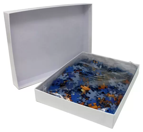
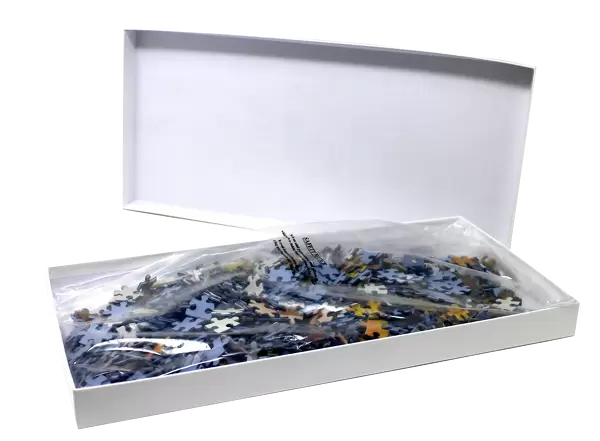
Jigsaw Puzzle (400 Pieces)
Discover the intricacies of nature with our Media Storehouse Jigsaw Puzzles! This captivating puzzle features a coloured Scanning Electron Micrograph (SEM) of human tooth enamel by Science Photo Library. Bring the mesmerizing world of science into your home with this puzzle, where each piece fits together to reveal the intricate details of this essential component of our teeth. Engage your mind, challenge your problem-solving skills, and unveil the beauty of enamel, one piece at a time.
400 piece puzzles are custom made in the UK and hand-finished on 100% recycled 1.5 mm millboard. There is a level of repetition in jigsaw shapes with each matching piece away from its pair. The completed puzzle measures 31x47cm and is delivered packaged in an attractive presentation box specially designed to fit most letter box slots
Jigsaw Puzzles are an ideal gift for any occasion
Estimated Product Size is 47.2cm x 31.5cm (18.6" x 12.4")
These are individually made so all sizes are approximate
Artwork printed orientated as per the preview above, with landscape (horizontal) or portrait (vertical) orientation to match the source image.
EDITORS COMMENTS
This print showcases the intricate beauty of human tooth enamel. In this coloured scanning electron micrograph (SEM), we are granted a close-up view of the highly mineralized material that covers the crown of a tooth. The crown, which is the visible part projecting into our oral cavity, is protected by this translucent and incredibly hard substance known as enamel. Upon closer examination, one can observe protoplasmic fibres of epithelial cells terminating in this section, appearing in a delicate shade of pink. Enamel itself consists of parallel rods or prisms made up mostly of calcium phosphate and calcium carbonate. These prisms are arranged perpendicular to the surface, contributing to both its strength and unique appearance. The level of detail captured in this image is truly remarkable thanks to its magnification capabilities. At 550 times magnification on a 6x7cm size print, it allows us to appreciate every minute aspect with awe-inspiring clarity. For those seeking an even more immersive experience, enlarging it further reveals astonishing intricacies at 1,480 times magnification on an 8x6ins print or an astounding 785 times magnification on a 10x7cm master size. This photograph not only highlights the fascinating anatomy and structure within our teeth but also serves as a testament to the wonders that lie hidden within our own bodies.
MADE IN THE UK
Safe Shipping with 30 Day Money Back Guarantee
FREE PERSONALISATION*
We are proud to offer a range of customisation features including Personalised Captions, Color Filters and Picture Zoom Tools
SECURE PAYMENTS
We happily accept a wide range of payment options so you can pay for the things you need in the way that is most convenient for you
* Options may vary by product and licensing agreement. Zoomed Pictures can be adjusted in the Basket.