
Neuroanatomy Collection
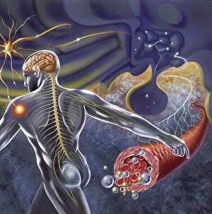
Neuroanatomy is a fascinating field that delves into the intricate structure and organization of the human brain


All Professionally Made to Order for Quick Shipping
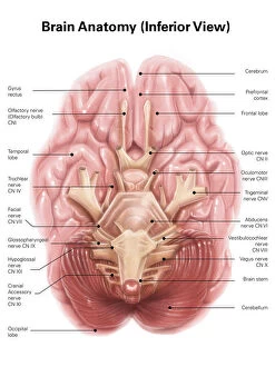
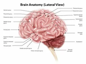
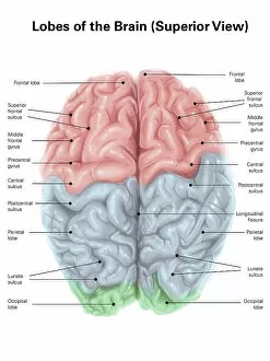
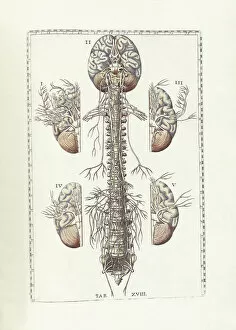
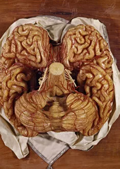
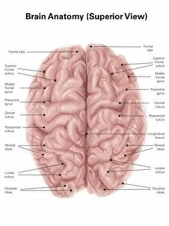
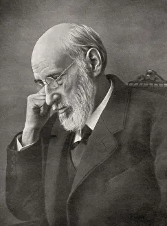
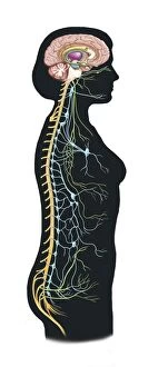
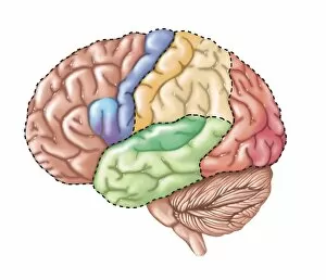
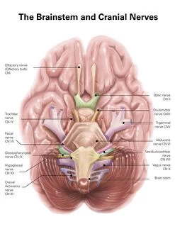
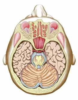
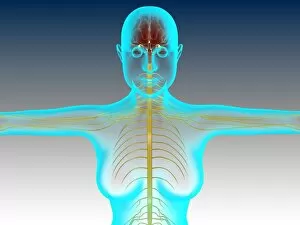
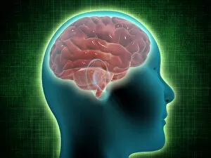
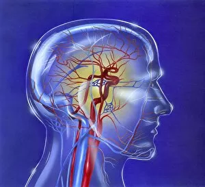
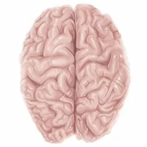
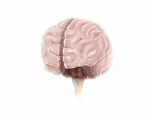
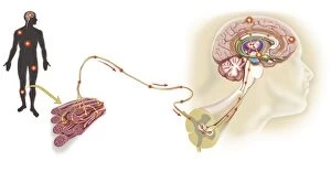
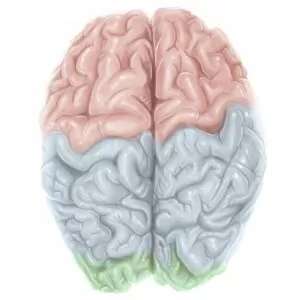
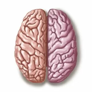
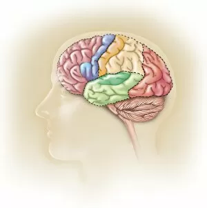
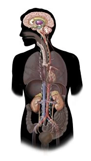
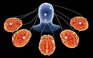
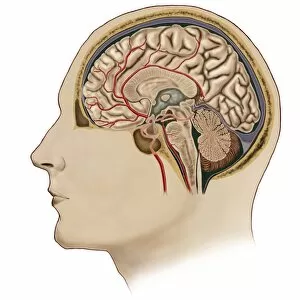
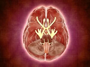
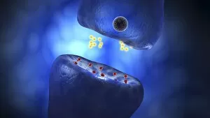
Neuroanatomy is a fascinating field that delves into the intricate structure and organization of the human brain. From its inferior view, we can observe the complex network of neurons and pathways that make up this remarkable organ. Moving to a superior view, colored lobes and labels help us identify different regions responsible for various functions such as cognition, emotion, and movement. The study of a rich history dating back centuries. Bartholomeo Eustachi's work on "The Science of Human Anatomy" laid the foundation for our understanding of brain anatomy. Another notable figure in this field is Franz Joseph Gall, whose chromolithograph captures his contributions as a German neuroanatomist and physiologist. Models like wax brains allow researchers to explore the intricacies in three dimensions, aiding their studies further. Santiago Ramon y Cajal's photograph from 1921 showcases his significant role as a Spanish neuroscientist and pathologist who made groundbreaking discoveries about neuronal structures. Understanding how the brain interacts with other systems is also crucial; an image displaying autonomic nervous system connections alongside limbic system components provides insight into these relationships. Additionally, labeled brain surface anatomy helps us navigate through different regions accurately. Lastly, let's not forget that both males and females possess unique nervous systems. A conceptual image highlighting female neural pathways reminds us that gender plays a role in neuroanatomy too. Neuroanatomy unravels the mysteries hidden within our minds by exploring its structure from multiple perspectives - inferior views revealing underlying complexity while superior views highlight functional specialization. Through historical figures like Eustachi, Gall, Cajal along with models and images showcasing various aspects of brain anatomy including connectivity patterns between systems or gender differences - it becomes clear why this discipline remains at the forefront of scientific exploration today.