
Glass Frame : Coloured SEM showing the ovulation process


![]()
Mounted Prints from Science Photo Library
Coloured SEM showing the ovulation process
Ovulation. Coloured scanning electron micrograph of the ovulation process. The egg (red at centre) has ruptured the external surface of the ovary (dark green), known as the germinal epithelium, and has started its journey through the Fallopian tube towards the uterus. The image clearly shows the thick glycoprotein layer (red), the zona pellucida, which entirely surrounds the egg. The egg is also partly surrounded by cells (granulosa cells light green) and fluid (liquor folliculi yellow), which formerly provided nutrients and protection to the egg in the ovary. Magnification: x1850 at 6x7cm size
Science Photo Library features Science and Medical images including photos and illustrations
Media ID 6454621
© PROF. P. MOTTA/DEPT. OF ANATOMY/UNIVERSITY LA SAPIENZA , ROME/SCIENCE PHOTO LIBRARY
Female Reproductive System Ovulation Ovum Re Production Zona Pellucida
7"x5" Glass Mount
Wall mounted or free-standing, these black edged glass frames feature a smooth chamfered edge and a stylish black border (on back face of the glass). Manufactured from 4mm thick glass, Glass Mounts are a durable, professional way of displaying and protecting your prints. Your 7x5 print is slotted into the back of the frame so can easily be changed if needed.
Tempered Glass Mounts are ideal for wall display, plus the smaller sizes can also be used free-standing via an integral stand
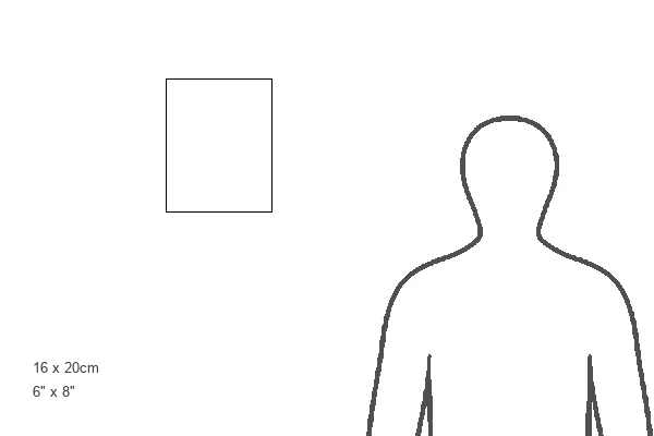
Estimated Image Size (if not cropped) is 12.7cm x 17.7cm (5" x 7")
Estimated Product Size is 16.2cm x 20.3cm (6.4" x 8")
These are individually made so all sizes are approximate
Artwork printed orientated as per the preview above, with portrait (vertical) orientation to match the source image.
EDITORS COMMENTS
This print showcases the intricate process of ovulation in stunning detail. In this coloured scanning electron micrograph, we witness the moment when an egg, represented by a vibrant red hue at the centre, ruptures through the external surface of the ovary. This outer layer, known as the germinal epithelium and depicted in dark green, marks the beginning of its remarkable journey towards the uterus through the Fallopian tube. The image also highlights a crucial component surrounding the egg - a thick glycoprotein layer called zona pellucida. This distinct red ring entirely envelops and protects the egg throughout its passage. Surrounding it are delicate granulosa cells depicted in light green that once provided nourishment and safeguarded this precious entity within its ovarian dwelling. Furthermore, we can observe liquor folliculi – a fluid shown in yellow – which played an essential role in nurturing and supporting this developing egg during its time within the ovary. With a magnification level of x1850 at 6x7cm size, this photograph offers us an extraordinary glimpse into one of nature's most miraculous processes: human ovulation. It serves as a testament to both our understanding of reproductive anatomy and our appreciation for life's intricate mechanisms.
MADE IN THE UK
Safe Shipping with 30 Day Money Back Guarantee
FREE PERSONALISATION*
We are proud to offer a range of customisation features including Personalised Captions, Color Filters and Picture Zoom Tools
SECURE PAYMENTS
We happily accept a wide range of payment options so you can pay for the things you need in the way that is most convenient for you
* Options may vary by product and licensing agreement. Zoomed Pictures can be adjusted in the Basket.