
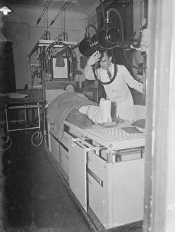
Xray Collection (page 16)
"Unveiling the Intricacies: Exploring the World Through X-ray Vision" Delving into the depths of our bodies


All Professionally Made to Order for Quick Shipping
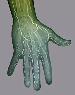
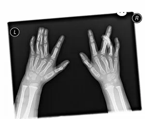
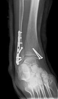
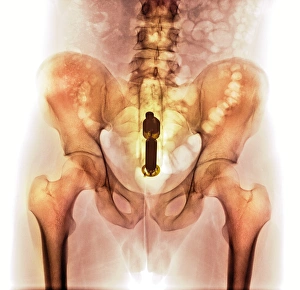
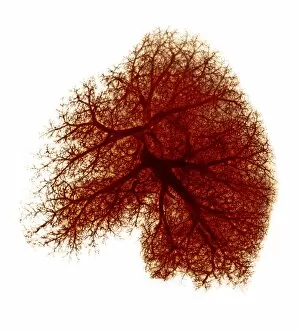
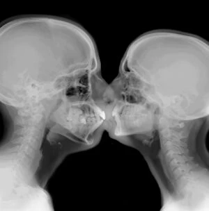
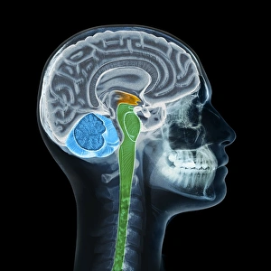
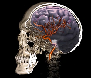
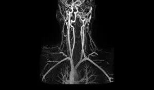
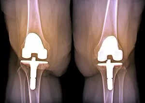
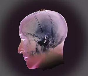
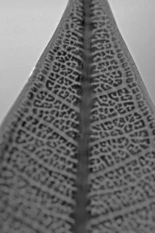
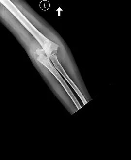
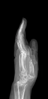
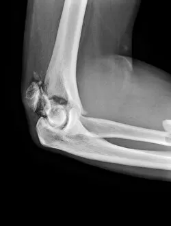
"Unveiling the Intricacies: Exploring the World Through X-ray Vision" Delving into the depths of our bodies, x-rays reveal the intricate network of brain blood vessels in a captivating 3D angiogram from 1981. A broken wrist bone captured in an x-ray, showcasing the resilience and fragility of our skeletal system. Hands outstretched under the penetrating gaze of an x-ray machine, revealing their hidden secrets and stories. The unexpected beauty found within an x-ray image of a stiletto high-heeled shoe, exposing its structural elegance from a unique perspective. An enchanting amaryllis flower brought to life through an ethereal x-ray, unraveling its delicate petals and mysterious allure. Witnessing artistry in motion as an MRI-style leg clad in a stiletto shoe is transformed into a mesmerizing x-ray masterpiece. A glimpse into perfect health with an immaculate knee showcased in all its glory through a crystal-clear x-ray image. Innocence encapsulated within an ordinary child's head as seen through the lens of an illuminating x-ray scan. Parsley leaves suspended mid-air like ghostly apparitions when exposed to the probing eyes of an x-ray machine - nature's hidden wonders revealed. Tulip bulbs radiate vibrant energy as they burst forth with life when viewed through this extraordinary tulipa sp. -inspired floral spectacle created by X-rays' colorful touch. The majestic gladiolus stands tall even under scrutiny, displaying its inner strength and grace when subjected to intense rays that penetrate deep beneath its surface layers. Behold. The Oriental Stargazer Lily unveils her true colors through this vividly colored X-Ray portrait.