Fraud Blocker Skip to main content

X Ray Machine Collection (page 4)

The incredible versatility of the x-ray machine never ceases to amaze us

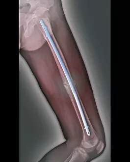
Background imageX Ray Machine Collection: Pinned thigh fracture, X-ray C016 / 6564

Pinned thigh fracture, X-ray C016 / 6564
Pinned thigh fracture. Coloured X-ray of the fractured femur (thigh bone) of a 52 year old patient. The broken bone has been fixed internally with a metal rod

Background imageX Ray Machine Collection: Scoliosis of the spine, X-ray C016 / 6565

Scoliosis of the spine, X-ray C016 / 6565
Scoliosis of the spine. Coloured X-ray of the chest of a 34 year old female patient with scoliosis (sideways curvature) of the spine

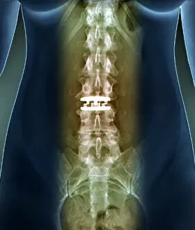
Background imageX Ray Machine Collection: Artificial spinal disc, X-ray C016 / 6561

Artificial spinal disc, X-ray C016 / 6561
Artificial spinal disc. Coloured X-ray of the spine of a 42 year old woman, showing a prosthetic spinal disc (white) in the lumbar (lower) spine

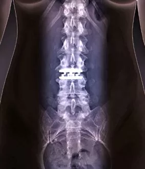
Background imageX Ray Machine Collection: Artificial spinal disc, X-ray C016 / 6562

Artificial spinal disc, X-ray C016 / 6562
Artificial spinal disc. Coloured X-ray of the spine of a 42 year old woman, showing a prosthetic spinal disc (white) in the lumbar (lower) spine

Background imageX Ray Machine Collection: Normal extended neck, X-ray C016 / 6424

Normal extended neck, X-ray C016 / 6424
Normal extended neck. Coloured X-ray of a 35 year old patients healthy extended cervical (neck) spine superimposed on a model of a human head

Background imageX Ray Machine Collection: Skull sutures, 3D CT scan C016 / 6403

Skull sutures, 3D CT scan C016 / 6403
Skull sutures. 3D computed tomography (CT) scan of the surface of a human skull, showing the sutures. Sutures are a type of rigid fibrous joint where the bones of the skull join

Background imageX Ray Machine Collection: Portal vein surgery, X-ray C016 / 6545

Portal vein surgery, X-ray C016 / 6545
Vein surgery. Coloured angiogram (X-ray) of surgery to lower the blood pressure in the portal vein of a 60 year old patient

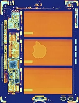
Background imageX Ray Machine Collection: Tablet computer, coloured x-ray C016 / 7194

Tablet computer, coloured x-ray C016 / 7194
Coloured x-ray of an iPad tablet computer showing the internal structure. The design of the tablet, the name iPad and the Apple logo are registered trademarks of Apple, Inc

Background imageX Ray Machine Collection: Portal vein surgery, X-ray C016 / 6544

Portal vein surgery, X-ray C016 / 6544
Vein surgery. Coloured angiogram (X-ray) of surgery to lower the blood pressure in the portal vein of a 60 year old patient

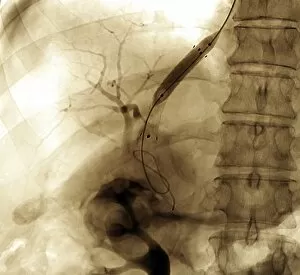
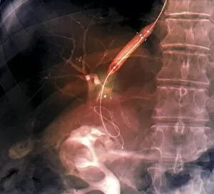
Background imageX Ray Machine Collection: Biliary drainage, X-ray C016 / 6559

Biliary drainage, X-ray C016 / 6559
Biliary drainage. Coloured X-ray of the abdomen of an 69 year old patient undergoing biliary drainage. This is a procedure to drain a blocked bile duct

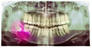
Background imageX Ray Machine Collection: Impacted wisdom tooth, panoral X-ray

Impacted wisdom tooth, panoral X-ray
Impacted wisdom tooth. Coloured panoral X-ray of the mouth of a patient with an impacted wisdom tooth (pink) in the right lower jaw

Background imageX Ray Machine Collection: Polycystic kidneys, CT scans C018 / 0571

Polycystic kidneys, CT scans C018 / 0571
Polycystic kidneys. Coloured computed tomography (CT) scans of the abdomen of a 28 year old patient with polycystic kidneys (pink)

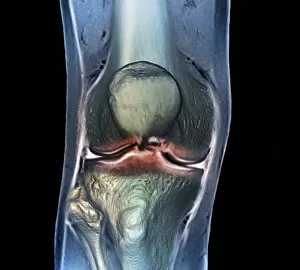
Background imageX Ray Machine Collection: Healthy knee, CT scan C018 / 0415

Healthy knee, CT scan C018 / 0415
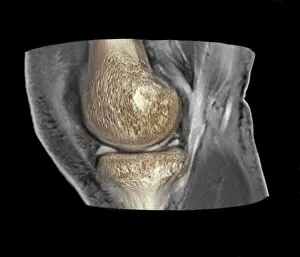
Healthy knee. Coloured frontal computed tomography (CT) scan projected on to a magnetic resonance imaging (MRI) scan of the knee of a 30 year old

Background imageX Ray Machine Collection: Polycystic kidneys, CT scans C018 / 0572

Polycystic kidneys, CT scans C018 / 0572
Polycystic kidneys. Coloured computed tomography (CT) scans of the abdomen of a 28 year old patient with polycystic kidneys (dark blue)

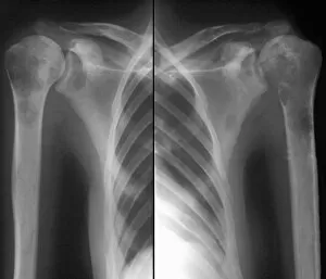
Background imageX Ray Machine Collection: Secondary bone cancer, X-ray C018 / 0594

Secondary bone cancer, X-ray C018 / 0594
Secondary bone cancer. X-ray of the shoulders of a 39 year old patient with metastatic (secondary) bone cancer. This cancer has spread from a primary thyroid cancer

Background imageX Ray Machine Collection: Trachea, 3D CT scan C018 / 0394

Trachea, 3D CT scan C018 / 0394
Trachea. Coloured 3D computed tomography (CT) scan of the healthy trachea (windpipe) of a 45 year old patient at the point that it bifurcates into the two primary bronchi (airways)

Background imageX Ray Machine Collection: Heart and lung disease, X-rays C018 / 0502

Heart and lung disease, X-rays C018 / 0502
Heart and lung disease. Coloured X-ray (top left) and computed tomography (CT) scans of the chest of an 80 year old patient with heart and lung disease

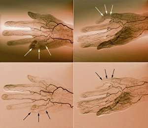
Background imageX Ray Machine Collection: Blood supply of the hand, angiograms C018 / 0389

Blood supply of the hand, angiograms C018 / 0389
Blood supply of the hand. Coloured angiograms (X-rays) of the blood vessels of the right hand of a 65 year old patient with Dupuytrens contracture

Background imageX Ray Machine Collection: Healthy knee, CT scan C018 / 0414

Healthy knee, CT scan C018 / 0414
Healthy knee. Coloured frontal computed tomography (CT) scan projected on to a magnetic resonance imaging (MRI) scan of the knee of a 30 year old

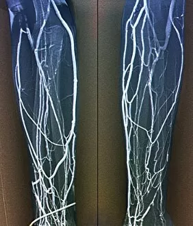
Background imageX Ray Machine Collection: Forearm veins, X-ray C018 / 0475

Forearm veins, X-ray C018 / 0475
Forearm veins. Coloured venogram (vein X-ray) of the veins in the forearms of a 43 year old patient in need of dialysis. This venogram has been taken to evaluate the health of the veins prior to

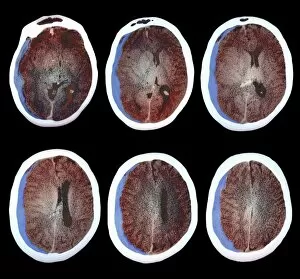
Background imageX Ray Machine Collection: Brain haemorrhage, CT scans C018 / 0569

Brain haemorrhage, CT scans C018 / 0569
Brain haemorrhage. Coloured computed tomography (CT) scans of the brain of a 49 year old patient with a subdural haematoma (blue)

Background imageX Ray Machine Collection: Aneurysm treatment, angiogram C018 / 0537

Aneurysm treatment, angiogram C018 / 0537
Aneurysm treatment. Coloured angiogram (X-ray) overlaid on a computer model of the treatment of a 39 year old patient with an aneurysm of the posterior communicating artery

Background imageX Ray Machine Collection: Voiding cystourethrogram, X-rays C018 / 0577

Voiding cystourethrogram, X-rays C018 / 0577
Voiding cystourethrogram. X-ray of a 54 year old male patient undergoing a voiding cystourethrogram. This procedure involves liquid containing a radio-opaque dye being placed in the bladder via a

Background imageX Ray Machine Collection: Knee injury, 3D CT scan C018 / 0648

Knee injury, 3D CT scan C018 / 0648
Knee injury. Coloured 3D computed tomography (CT) scan of a knee with a meniscal tear (white line at right between bones)

Background imageX Ray Machine Collection: Voiding cystourethrogram, X-rays C018 / 0578

Voiding cystourethrogram, X-rays C018 / 0578
Voiding cystourethrogram. Coloured X-ray of a 54 year old male patient undergoing a voiding cystourethrogram. This procedure involves liquid containing a radio-opaque dye being placed in the bladder

Background imageX Ray Machine Collection: Secondary bone cancer, X-ray C018 / 0531

Secondary bone cancer, X-ray C018 / 0531
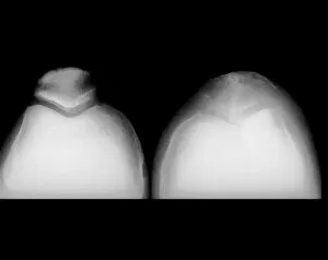
Secondary bone cancer. X-ray of the knees of a 68 year old patient with metastatic (secondary) bone cancer of the left patella (knee cap). This cancer has spread from a primary lung cancer

Background imageX Ray Machine Collection: 17 month old babys pelvis, X-ray

17 month old babys pelvis, X-ray
Seventeen month old babys pelvis, coloured X-ray. The hip joints (centre right and centre left) are still forming

Background imageX Ray Machine Collection: Aneurysm treatment, angiogram C018 / 0536

Aneurysm treatment, angiogram C018 / 0536
Aneurysm treatment. Angiogram (X-ray) overlaid on a computer model of the treatment of a 39 year old patient with an aneurysm of the posterior communicating artery

Background imageX Ray Machine Collection: Osteoarthritis of the hip, X-ray C018 / 0593

Osteoarthritis of the hip, X-ray C018 / 0593
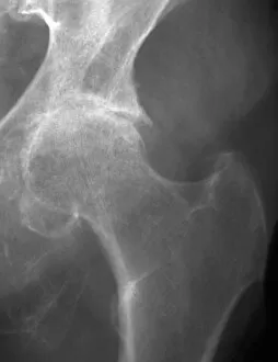
Osteoarthritis of the hip. X-ray the left hip of a 74 year old patient showing osteoarthritis. The ball at the top of the femur (thigh bone) fits into the socket of the hip

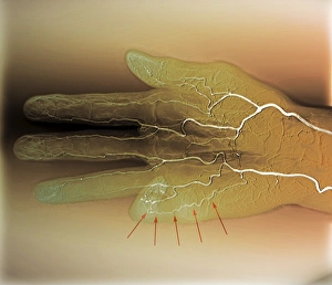
Background imageX Ray Machine Collection: Blood supply of the hand, angiogram C018 / 0387

Blood supply of the hand, angiogram C018 / 0387
Blood supply of the hand. Coloured angiogram (X-ray) of the blood vessels of the right hand of a 65 year old patient with Dupuytrens contracture
Background imageX Ray Machine Collection: Knee injury, 3D CT scan C018 / 0639

Knee injury, 3D CT scan C018 / 0639
Knee injury. Coloured 3D computed tomography (CT) scan of a knee with a meniscal tear (indicated by arrows). The menisci are fibrocartilage crescents that act as shock absorbers between the femur

Background imageX Ray Machine Collection: Hydrocephalus, CT scan C018 / 0568

Hydrocephalus, CT scan C018 / 0568
Hydrocephalus. Coloured computed tomography (CT) scan of the brain of a 22 year old patient with hydrocephalus (fluid in the brain)

Background imageX Ray Machine Collection: Liver cancer, CT scan C018 / 0461

Liver cancer, CT scan C018 / 0461
Liver cancer. Coloured computed tomography (CT) scan of the abdomen of a 50 year old alcoholic patient with liver cancer. The liver (orange) is enlarged due to the presence of the tumour

Background imageX Ray Machine Collection: Fractured skull, X-ray C018 / 0591

Fractured skull, X-ray C018 / 0591
Fractured skull, X-ray. The fracture is the grey line at right that splits into two near the temple

Background imageX Ray Machine Collection: Jeune syndrome, X-ray C018 / 0410

Jeune syndrome, X-ray C018 / 0410
Jeune syndrome. X-ray of the chest of a 2 year old with Jeune syndrome, also known as asphyxiating thoracic dystrophy. This inherited disease affects the development of the bones and cartilage

Background imageX Ray Machine Collection: Arthritic hands, X-ray C018 / 0534

Arthritic hands, X-ray C018 / 0534
Arthritic hands. Coloured X-ray of the hands of a 72 year old patient with rheumatoid arthritis. The wrist and finger joints have become inflamed and swollen

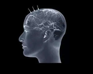
Background imageX Ray Machine Collection: Thrombophlebitis, angiogram C018 / 0529

Thrombophlebitis, angiogram C018 / 0529
Thrombophlebitis. Coloured angiogram (X-ray) of the brain of a 28 year old female patient who had thrombophelibitis of the superior sagittal sinus (indicated by arrows)

Background imageX Ray Machine Collection: Knee injury, 3D CT scan C018 / 0645

Knee injury, 3D CT scan C018 / 0645
Knee injury. Coloured 3D computed tomography (CT) scan of a knee with a meniscal tear (blue line at right between bones). The menisci are fibrocartilage crescents that act as shock absorbers between

Background imageX Ray Machine Collection: Secondary bone cancer, X-ray C018 / 0595

Secondary bone cancer, X-ray C018 / 0595
Secondary bone cancer. X-ray of the left shoulder of a 39 year old patient with metastatic (secondary) bone cancer. This cancer has spread from a primary thyroid cancer

Background imageX Ray Machine Collection: Hydropneumothorax, X-ray C018 / 0586

Hydropneumothorax, X-ray C018 / 0586
Hydropneumothorax. Coloured X-ray of the chest of a 62 year old patient with a hydropneumothorax of the left lung (dark area at upper centre)

Background imageX Ray Machine Collection: Colon cancer, barium X-ray C018 / 0574

Colon cancer, barium X-ray C018 / 0574
Colon cancer. Coloured X-ray of the large intestine of a 62 year old patient with cancer of the sigmoid colon. The patient was given a barium enema prior to the X-ray to show up any abnormalities

Background imageX Ray Machine Collection: Liver cancer, CT scan C018 / 0462

Liver cancer, CT scan C018 / 0462
Liver cancer. Computed tomography (CT) scan of the abdomen of a 50 year old alcoholic patient with liver cancer. The liver (left to upper centre) is enlarged due to the presence of the tumour

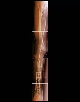
Background imageX Ray Machine Collection: Pinned broken leg, angiogram C018 / 0392

Pinned broken leg, angiogram C018 / 0392
Pinned broken leg. Coloured angiogram of the right leg of a 22 year old patient that sustained an open tibial (shin bone) fracture in a motorcycle accident. The fracture has been aligned and pinned

Background imageX Ray Machine Collection: Trachea, 3D CT scan C018 / 0393

Trachea, 3D CT scan C018 / 0393
Trachea. Coloured 3D computed tomography (CT) scan of the healthy trachea (windpipe) of a 45 year old patient at the point that it bifurcates into the two primary bronchi (airways)

Background imageX Ray Machine Collection: Liver cancer, CT scan C018 / 0460

Liver cancer, CT scan C018 / 0460
Liver cancer. Coloured computed tomography (CT) scan of the abdomen of a 50 year old alcoholic patient with liver cancer. The liver (orange) is enlarged due to the presence of the tumour

Background imageX Ray Machine Collection: Aneurysm treatment, angiogram C018 / 0525

Aneurysm treatment, angiogram C018 / 0525
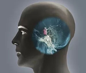
Aneurysm treatment. Coloured angiogram (X-ray) overlaid on a computer model of the treatment of a patient with an aneurysm of the internal carotid artery

Background imageX Ray Machine Collection: Arthritic hands, X-ray C018 / 0532

Arthritic hands, X-ray C018 / 0532
Arthritic hands. X-ray of the hands of a 72 year old patient with rheumatoid arthritis. The wrist and finger joints have become inflamed and swollen

Background imageX Ray Machine Collection: Hydrocephalus, CT scan C018 / 0567

Hydrocephalus, CT scan C018 / 0567
Hydrocephalus. Coloured computed tomography (CT) scan of the brain of a 22 year old patient with hydrocephalus (fluid in the brain)


All Professionally Made to Order for Quick Shipping

The incredible versatility of the x-ray machine never ceases to amaze us. From capturing intricate details of brain blood vessels in a 3D angiogram C007 / 1981, to diagnosing a broken wrist bone in X-ray C017 / 7187, this technology has revolutionized medical imaging. But it doesn't stop there. The x-ray machine has even ventured into the animal kingdom, revealing fascinating insights such as a horse's skull or the delicate skeleton of a seahorse. It seems that no creature is too small or too large for this remarkable tool. In addition to its medical applications, the x-ray machine also showcases its playful side. Who would have thought that skeletons could engage in sports? A rugby-playing skeleton and even one enjoying a refreshing drink are just some how this technology can bring humor and creativity to our lives. Not limited to humans and animals, the x-ray machine extends its reach into other fields as well. Panoramic dental X-rays help dentists assess oral health with precision while an individual holding a camera reveals how this invention itself can be captured on film. Furthermore, we witness how the x-ray machine aids in surgical procedures like total hip replacements by providing surgeons with detailed images for precise placement and alignment. Last but not least, let's not forget about exploring what lies beneath our own skin - normal skulls are examined through X-rays allowing us to marvel at the intricacies hidden within each one of us. From healthcare advancements to artistic expressions and scientific discoveries, these glimpses into various aspects of life demonstrate why the x-ray machine remains an invaluable tool that continues to shape our understanding of both human anatomy and beyond.