Fraud Blocker Skip to main content

X Ray Machine Collection (page 11)

The incredible versatility of the x-ray machine never ceases to amaze us

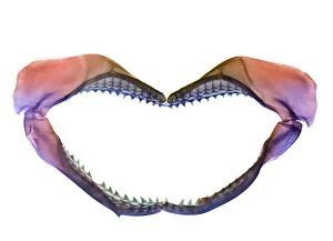
Background imageX Ray Machine Collection: Shark jaws

Shark jaws, coloured X-ray

Background imageX Ray Machine Collection: Jaws of a shark

Jaws of a shark, coloured X-ray

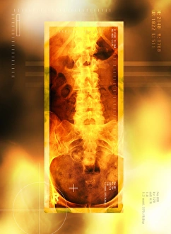
Background imageX Ray Machine Collection: Healthy lower back, X-ray

Healthy lower back, X-ray
Healthy lower back, coloured X-ray. The spine runs down centre. It is made up of vertebrae, block- shaped bones that support and protect the spinal cord (not seen)

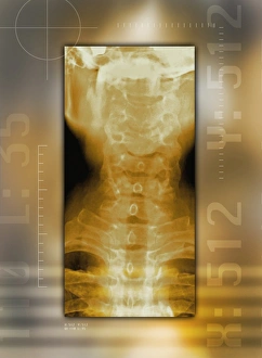
Background imageX Ray Machine Collection: Healthy neck bones, X-ray

Healthy neck bones, X-ray
Healthy neck bones, coloured X-ray. The neck contains seven vertebrae, the blocky bones that make up the spinal column (down centre)

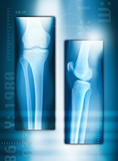
Background imageX Ray Machine Collection: Knee joint from front and side, X-ray

Knee joint from front and side, X-ray
Knee joint from the front (left) and side (right), coloured X-rays. The knee is the largest joint in the human body. It is formed from the articulation of the thigh bone (femur)

Background imageX Ray Machine Collection: Skeleton walking a marmoset, X-ray

Skeleton walking a marmoset, X-ray
Walking a pet marmoset, conceptual X-ray. The marmoset X-ray is not to scale: marmosets reach a maximum length of around 20 centimetres

Background imageX Ray Machine Collection: Skeletons of a human and rats, X-ray

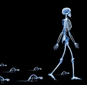
Skeletons of a human and rats, X-ray
Human and rat skeletons, conceptual X-ray. This recalls the legend of the Pied Piper, who drew the rats from the town of Hamelin, Germany, in the Middle Ages

Background imageX Ray Machine Collection: Technological dominance, conceptual image

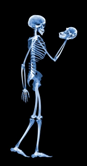
Technological dominance, conceptual image
Evolution. Conceptual X-ray image of a human skeleton holding the skull of a chimpanzee (Pan troglodytes), the closest living relative of humans

Background imageX Ray Machine Collection: Skeleton smoking

Skeleton smoking
Skeleton. Computer-enhanced X-ray of a side view of a skeleton smoking a cigarette whilst walking

Background imageX Ray Machine Collection: Skeleton using a mobile phone

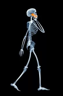
Skeleton using a mobile phone
Skeleton. Computer-enhanced X-ray of a side view of a skeleton using a mobile phone whilst walking

Background imageX Ray Machine Collection: Dolphin X-ray

Dolphin X-ray. Technician examining a digital X- ray of a bottlenose dolphin (Tursiops truncatus), Navy Marine Mammal Program (NMMP), San Diego, USA

Background imageX Ray Machine Collection: Woman with neck X-ray

Woman with neck X-ray
MODEL RELEASED. Woman and X-ray. Composite image of a nude woman and an X-ray of her neck in front view (left) and side view (right). This may represent worrying about the results of a scan

Background imageX Ray Machine Collection: High resolution X-ray CT scanner

High resolution X-ray CT scanner
High resolution X-ray computed tomography (CT) scanner, University of Texas, Austin, USA. This scanner is used by scientists studying rocks, fossils and biological specimens

Background imageX Ray Machine Collection: Back pain due to poor driving position

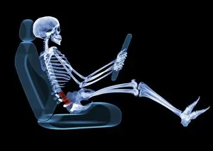
Back pain due to poor driving position
Lower back pain (red) caused by an improper sitting position while driving. This X-ray shows the bones of the lower back to be curved rather than straight

Background imageX Ray Machine Collection: Arthritic knee

Arthritic knee
Arthritis. Computer artwork of an X-ray of an arthritic knee

Background imageX Ray Machine Collection: Osteoporosis of the back, X-ray

Osteoporosis of the back, X-ray
Osteoporosis of the back, coloured X-ray. Osteoporosis is a condition in which bone density is reduced, weakening the bones and making them susceptible to fractures

Background imageX Ray Machine Collection: Osteoporotic spine, X-ray

Osteoporotic spine, X-ray
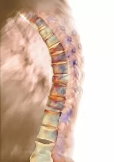
Osteoporotic spine, coloured X-ray. The spine is seen in side view, with the front of the body at right. It is seen to be curved, bowing out from top right to centre left then back right again

Background imageX Ray Machine Collection: Fractured elbow, X-rays

Fractured elbow, X-rays
Fractured elbow, coloured X-ray. The elbow is seen straight (left) and bent (right). In the bent view, a small patch of bone is seen above the joint

Background imageX Ray Machine Collection: Healing ankle fracture, X-ray

Healing ankle fracture, X-ray
Healing ankle fracture, coloured X-rays. The ankle is seen from the front at left and from the side at right. The break is at the bottom of the fibula (left in front view)

Background imageX Ray Machine Collection: Fractured shoulder, X-rays

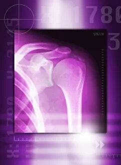
Fractured shoulder, X-rays
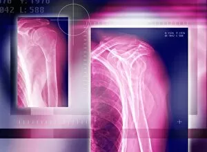
Shoulder fracture, coloured X-rays. The joint is seen from the side at left and from the front at right. The joint has not been dislocated by the fracture

Background imageX Ray Machine Collection: Broken collar bone, X-ray

Broken collar bone, X-ray
Broken collar bone. Coloured frontal X-ray of a patient with a fractured left collar bone (clavicle). The fracture is at the top right of the shoulder

Background imageX Ray Machine Collection: Knee effusion

Knee effusion. Coloured X-ray of the swelling of an injured knee caused by effusion. Effusion is a painful condition whereby fluid accumulates in the joint space, limiting movement

Background imageX Ray Machine Collection: Narrowed arteries, X-ray

Narrowed arteries, X-ray
Narrowed arteries. Coloured fluoroscope (X-ray) of the abdomen of a patient with stenosis (narrowing, purple areas) of the coeliac trunk (top) and the superior mesenteric artery (SMA, bottom)

Background imageX Ray Machine Collection: Arterial stents, X-ray

Arterial stents, X-ray
Arterial stents. Coloured X-ray of the abdomen of a patient with stents (wire mesh tubes) in two arteries. The aorta, the bodys main artery is running down centre

Background imageX Ray Machine Collection: Staghorn kidney stone, X-ray

Staghorn kidney stone, X-ray
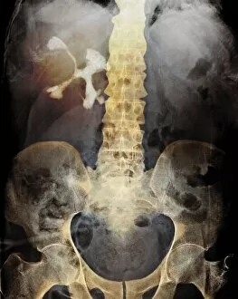
Staghorn kidney stone. Coloured X-ray of the abdomen of a patient with a large staghorn stone (white, upper left) in the right kidney

Background imageX Ray Machine Collection: Gases in the abdomen, X-ray

Gases in the abdomen, X-ray
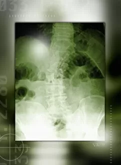
Bloated abdomen in tympanites, coloured X-ray. Tympanites is swelling of the abdomen due to the accumulation of gases in the intestines or the peritoneal cavity. Gas pockets are seen as dark spaces

Background imageX Ray Machine Collection: Spinal osteoporosis, X-ray

Spinal osteoporosis, X-ray
Spinal osteoporosis. Coloured X-ray of the lumbar region of the spine of an elderly female patient with osteoporosis. The bones have lost their density and have become porous and fragile

Background imageX Ray Machine Collection: Heart failure, chest X-ray

Heart failure, chest X-ray
Heart failure, or cardiomyopathy, coloured chest X-ray. The heart (pale orange, lower right) is greatly enlarged (ventricular hypertrophy)

Background imageX Ray Machine Collection: Enlarged heart, X-ray

Enlarged heart, X-ray
Enlarged heart. Coloured X-ray showing a patient with left ventricular hypertrophy (LVH), or enlargement of the left ventricle (lower chamber)

Background imageX Ray Machine Collection: Fibrosis of the lung, X-ray

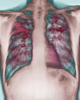
Fibrosis of the lung, X-ray
Lung fibrosis. Coloured X-ray of the chest of a patient with pulmonary (lung) fibrosis, or interstitial lung disease. Pulmonary fibrosis is the overgrowth of fibrous scar tissue (dark red)

Background imageX Ray Machine Collection: Calcified shoulder joint, X-ray

Calcified shoulder joint, X-ray
Calcified shoulder joint. Coloured X-ray of a calcified shoulder joint. There is a deposit of calcium at the top right of the shoulder joint

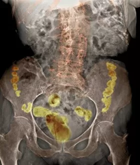
Background imageX Ray Machine Collection: Crohns disease, X-ray

Crohns disease, X-ray

Background imageX Ray Machine Collection: Bronchitis, X-ray

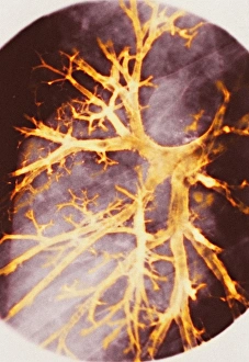
Bronchitis, X-ray
Bronchitis. Coloured bronchography (X-ray) of swollen bronchioles (orange) in the right lung. Bronchitis causes the network of airways in the lungs to become swollen and produce mucus

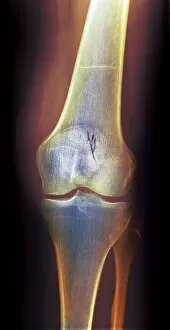
Background imageX Ray Machine Collection: Arthritic knee, X-ray

Arthritic knee, X-ray
Arthritic knee. Coloured X-ray of the knee of a patient with rheumatoid arthritis. At top is the femur (thigh bone), at bottom is the tibia (shinbone), with the smaller fibula to its right

Background imageX Ray Machine Collection: Electric train, X-ray

Electric train, X-ray
Electric train. X-ray showing the mechanism inside an electric toy train

Background imageX Ray Machine Collection: Toy tin tractor, X-ray

Toy tin tractor, X-ray
Toy tin tractor. X-ray showing the mechanism inside a toy tractor

Background imageX Ray Machine Collection: Neck X-ray

Neck X-ray
MODEL RELEASED. Neck X-ray

Background imageX Ray Machine Collection: Arm X-ray

Arm X-ray
MODEL RELEASED. Arm X-ray

Background imageX Ray Machine Collection: Pelvic X-ray

Pelvic X-ray
MODEL RELEASED. Pelvic X-ray

Background imageX Ray Machine Collection: Normal lower jaw, X-ray

Normal lower jaw, X-ray
Normal lower jaw. Coloured profile X-ray of a human mandible (lower jaw). This is attached to the skull (top) by the temporomandibular joint (TMJ, upper left)

Background imageX Ray Machine Collection: Amputated toe, X-ray

Amputated toe, X-ray
Amputated toe. Coloured frontal (left) and oblique (right) X-rays of a foot of a diabetic, showing an amputated second toe

Background imageX Ray Machine Collection: Normal leg veins, angiogram

Normal leg veins, angiogram
Normal leg veins, coloured angiogram (blood vessel X-ray)

Background imageX Ray Machine Collection: Arthrosis of the knee, X-ray

Arthrosis of the knee, X-ray
Arthrosis of the knee, coloured frontal X-ray. The joint is swollen and there are osteophytes, abnormal bony growths (pointed protrusions), seen on the bone at centre

Background imageX Ray Machine Collection: Normal brain, CT scans

Normal brain, CT scans
Normal brain. Coloured axial (cross-sectional) computed tomography (CT) scans of a healthy human head. The front of the head is at right in each scan

Background imageX Ray Machine Collection: Normal brain, CT scan

Normal brain, CT scan
Normal brain, coloured axial (cross-sectional) computed tomography (CT) scan. The front of the head is at top. The brain is yellow (lower centre)

Background imageX Ray Machine Collection: Normal lower leg, X-ray

Normal lower leg, X-ray
Normal lower leg, coloured frontal X-ray. The tibia (shinbone, left) and the smaller fibula (centre) articulate with the foot bones (bottom) to form the ankle joint

Background imageX Ray Machine Collection: Pinned foot bone fracture, X-ray

Pinned foot bone fracture, X-ray
Pinned foot bone fracture. Coloured X-rays of the right foot, showing a metal plate and screws (white) in the foot bone beneath the little toe (upper right)

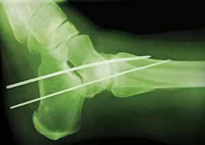
Background imageX Ray Machine Collection: Temporary ankle immobilisation, X-ray

Temporary ankle immobilisation, X-ray
Temporary ankle immobilisation, coloured profile X-ray. These long pins (white rods) are holding the bone fragments in position until they can be operated on and reset properly


All Professionally Made to Order for Quick Shipping

The incredible versatility of the x-ray machine never ceases to amaze us. From capturing intricate details of brain blood vessels in a 3D angiogram C007 / 1981, to diagnosing a broken wrist bone in X-ray C017 / 7187, this technology has revolutionized medical imaging. But it doesn't stop there. The x-ray machine has even ventured into the animal kingdom, revealing fascinating insights such as a horse's skull or the delicate skeleton of a seahorse. It seems that no creature is too small or too large for this remarkable tool. In addition to its medical applications, the x-ray machine also showcases its playful side. Who would have thought that skeletons could engage in sports? A rugby-playing skeleton and even one enjoying a refreshing drink are just some how this technology can bring humor and creativity to our lives. Not limited to humans and animals, the x-ray machine extends its reach into other fields as well. Panoramic dental X-rays help dentists assess oral health with precision while an individual holding a camera reveals how this invention itself can be captured on film. Furthermore, we witness how the x-ray machine aids in surgical procedures like total hip replacements by providing surgeons with detailed images for precise placement and alignment. Last but not least, let's not forget about exploring what lies beneath our own skin - normal skulls are examined through X-rays allowing us to marvel at the intricacies hidden within each one of us. From healthcare advancements to artistic expressions and scientific discoveries, these glimpses into various aspects of life demonstrate why the x-ray machine remains an invaluable tool that continues to shape our understanding of both human anatomy and beyond.