
Trabeculae Collection
"Trabeculae: The Intricate Network of Bone Structure Unveiled" Discover the mesmerizing beauty of trabeculae, as seen in artwork C016/7504


All Professionally Made to Order for Quick Shipping
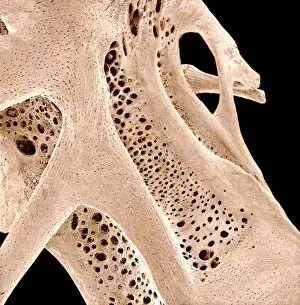
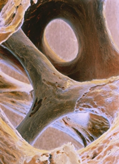
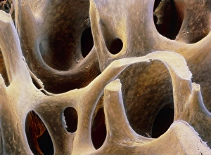
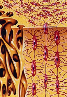
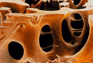
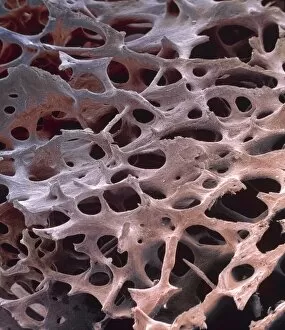
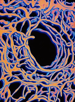
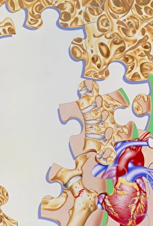
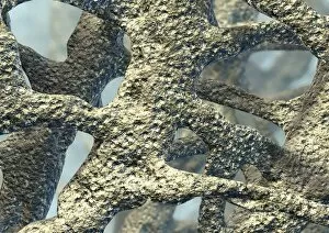
"Trabeculae: The Intricate Network of Bone Structure Unveiled" Discover the mesmerizing beauty of trabeculae, as seen in artwork C016/7504. These delicate bone structures form a captivating pattern that supports and strengthens our skeletal system. Intricately woven like an artistic masterpiece, it can visible through SEM images showcasing bone tissue (SEM C013/4768). These microscopic marvels reveal the intricate architecture within our bones, providing insight into their strength and resilience. Not limited to human bones alone, fish bones also exhibit this fascinating network. Delve into the underwater world with SEM images of fish bone (SEM C015/8068) where trabeculae create a framework for support and flexibility. The thigh bone head X-ray offers a glimpse into how trabeculae extend beyond mere aesthetics. They play a vital role in maintaining healthy joints by distributing forces evenly across the bone surface. As we explore further, artwork such as C016/7545 showcases another dimension of trabecular beauty. Its abstract representation reminds us that even within scientific realms, art can be found. Beyond bones, lymph nodes also possess their own unique arrangement of trabeculae. Admire these intricate pathways through artwork C013/4632 and C013/4631 as they guide immune cells on their mission to protect our bodies from harm. Whether observed under an electron microscope or portrayed artistically, trabeculae never fail to captivate us with their elegance and functionality. Let us appreciate these hidden wonders that make up the foundation of life itself – reminding us that true beauty lies not only on the surface but deep within our very being.