Fraud Blocker Skip to main content

Tarsals Collection

Tarsals, the foundation of our feet

Background imageTarsals Collection: Normal foot, X-ray

Normal foot, X-ray
Normal foot. Coloured X-ray of a healthy human foot seen from the top (antero-posterior or dorso- plantar projection). The foot comprises 26 bones and two sesamoid bones (not seen)

Background imageTarsals Collection: Skeleton from below, X-ray artwork

Skeleton from below, X-ray artwork
Skeleton. Computer x-ray artwork of a human skeleton viewed from below

Background imageTarsals Collection: Outer ankle ligaments, artwork C013 / 4452

Outer ankle ligaments, artwork C013 / 4452
Outer ankle ligaments. Computer artwork of the bones and ligaments (white) of the right foot and ankle seen from the side

Background imageTarsals Collection: Inner ankle ligaments, artwork C013 / 4451

Inner ankle ligaments, artwork C013 / 4451
Inner ankle ligaments. Computer artwork of the bones and ligaments (white) of the right foot and ankle seen from the side

Background imageTarsals Collection: Childs foot, X-ray

Childs foot, X-ray
Childs foot. Coloured X-ray of the healthy right foot of an eight year old boy. The toes are made up of the phalanx bones (phalanges), two for the big toe (lower right) and three for the others

Background imageTarsals Collection: Pinned foot bones after surgery, X-ray

Pinned foot bones after surgery, X-ray
Pinned foot bones after surgery. Lateral (side view) X-ray showing pins (white) inserted into the bones of a patients foot

Background imageTarsals Collection: Leg ligaments, artwork C013 / 4656

Leg ligaments, artwork C013 / 4656
Leg anatomy. Computer artwork of an anterior view of a human leg, showing the muscles and tendons (brown and white), bones and ligaments (white), arteries and veins (red and blue), nerves (yellow)

Background imageTarsals Collection: Outer ankle ligaments, artwork C013 / 4456

Outer ankle ligaments, artwork C013 / 4456
Outer ankle ligaments. Computer artwork of the bones and ligaments (white) of the feet and ankles from an oblique side view, with the outer side of the right foot at left

Background imageTarsals Collection: Outer ankle ligaments, artwork C013 / 4455

Outer ankle ligaments, artwork C013 / 4455
Outer ankle ligaments. Computer artwork of the bones and ligaments (white) of the feet and ankles from an oblique side view, with the outer side of the left foot at upper centre

Background imageTarsals Collection: Foot bones

Foot bones, computer artwork. The toes are made up of the phalanx bones (phalanges), two for the big toe (lower left) and three for the others

Background imageTarsals Collection: Ankle bones and ligaments

Ankle bones and ligaments. Historical anatomical artwork of ankle bones (yellow) and ligaments (pale blue). Ligaments are bands of fibrous tissue that hold bones together at their joints

Background imageTarsals Collection: Flat foot, X-ray

Flat foot, X-ray
Coloured X-ray (side view) of the flat foot of a standing patient. The decreased arching of the foot in flat foot (pes planus) means the sole lies flat on the ground and the heel bone

Background imageTarsals Collection: Foot fork-stabbing injury, X-ray

Foot fork-stabbing injury, X-ray
Foot fork-stabbing injury, coloured X-ray. The fork (white, top) has been stabbed into the area of the foot where the metatarsals join the tarsal bones

Background imageTarsals Collection: Skeletons feet, artwork

Skeletons feet, artwork
Skeletons feet. Computer artwork showing the bones of the feet of a human skeleton that is lying down


All Professionally Made to Order for Quick Shipping

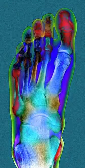
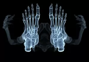
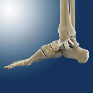
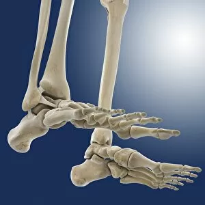
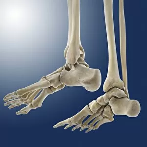
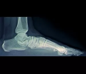
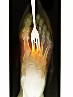
Tarsals, the foundation of our feet. 🦶🏻✨ From a normal foot X-ray to intricate artwork depicting the skeleton from below, these small yet crucial bones play a significant role in our mobility. In an X-ray artwork showcasing tarsals, we witness their placement within the foot's structure and how they contribute to its overall functionality. The outer ankle ligaments (artwork C013 / 4452) and inner ankle ligaments (artwork C013 / 4451) further emphasize their importance by highlighting the connections that keep everything in place. Even in a child's foot X-ray or after surgery with pinned foot bones, tarsals remain steadfast as key components for stability and movement. Their presence is beautifully captured in detailed artwork representing individual foot bones. But it doesn't stop there. Ligaments also come into play when discussing tarsals' significance. Artwork C013 / 4656 showcases leg ligaments while outer ankle ligament artworks (C013 / 4456 & C013 / 4455) provide insight into how tarsals interact with other structures. Ultimately, whether through X-rays or artistic representations, studying tarsals allows us to appreciate the complexity of our feet's anatomy. So next time you take a step forward, remember that these remarkable bones are working tirelessly beneath your skin to support every move you make.