
Superficial Collection
"Unveiling the Superficial: Exploring Art, Anatomy, and Society in Captivating Images" Step into a world where art meets anatomy


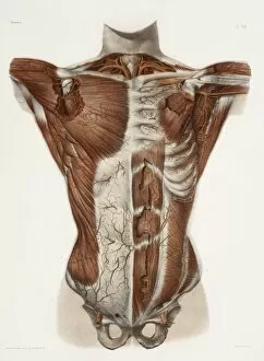


All Professionally Made to Order for Quick Shipping
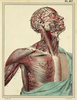
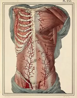
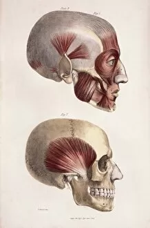
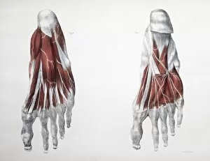
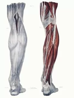
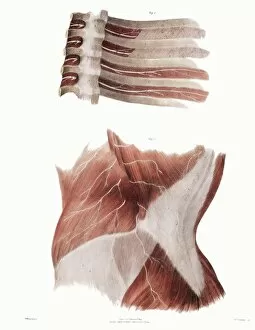
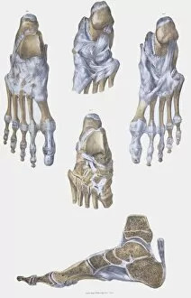
"Unveiling the Superficial: Exploring Art, Anatomy, and Society in Captivating Images" Step into a world where art meets anatomy, and society's superficiality is laid bare. From intricate 1825 artwork depicting head and chest arteries to mesmerizing illustrations of head and neck anatomy from 1831, these captivating visuals offer a glimpse into the intricacies of our bodies. Delve deeper as you explore face and neck muscles in an 1831 artwork or unravel the mysteries of cervical spinal nerves through a stunning 1844 illustration. These masterpieces not only showcase scientific knowledge but also highlight the artistic brilliance that brings them to life. But it's not just science that captivates us; society plays its part too. Witness a snapshot from an audience at a "coloured party, " transporting you back to another era when social gatherings were segregated yet filled with vibrancy. Marvel at architectural wonders created by T E Collcutt and Stanley Hamp, architects whose lithographs transport us to grand structures that stand tall even today. And don't miss out on fashion plates like "Elegant Woman with her doggie" from Galerie des Modes et Costumes - showcasing timeless elegance across generations. As we journey further, let your eyes wander over engravings displaying muscles of the chest and front arm while black-and-white photographs take you back in time to Cape Town's Feather Market - capturing moments frozen forever. Dive deep into anatomical engravings revealing the intricate network of lymphatics and glands on our heads, faces, and necks. Finally, witness post-mortem inner arm dissections providing insights into human anatomy beyond what meets the eye. In this collection spanning centuries past, we uncover both beauty and complexity - reminding us that beneath every superficial layer lies stories waiting to be discovered.