Fraud Blocker Skip to main content

Spiracle Collection

"Discovering the intricate beauty of spiracles in nature's creatures - from the mesmerizing eye

Background imageSpiracle Collection: Eye and spiracle detail of a Bluespotted / Ribbontail Stingray (Taeniura lymma) Sipadan Island

Eye and spiracle detail of a Bluespotted / Ribbontail Stingray (Taeniura lymma) Sipadan Island, Sabah, Borneo, Malaysia. May

Background imageSpiracle Collection: Mustelus canis, Smooth Dogfish, side view

Mustelus canis, Smooth Dogfish, side view

Background imageSpiracle Collection: Fruit fly spiracle, SEM

Fruit fly spiracle, SEM
Fruit fly (Drosophila melanogaster) spiracle. Coloured scanning electron micrograph (SEM) of the spiracle (air pore) of a male fruit fly

Background imageSpiracle Collection: Ant spiracle, SEM C018 / 0551

Ant spiracle, SEM C018 / 0551
Ant spiracle, coloured scanning electron micrograph (SEM). Spiracles are openings in an insects exoskeleton that connect to the tracheal tubes which deliver oxygen directly to tissues within

Background imageSpiracle Collection: Anopheles stephensi mosquito larva

Anopheles stephensi mosquito larva. The head is at left, the tail at right. The adult Anopheles stephensi female is a known vector for the tropical disease malaria

Background imageSpiracle Collection: Culex mosquito larva C017 / 8352

Culex mosquito larva C017 / 8352
Culex mosquito larva, with a breathing tube, on the waters surface. Mosquito larvae have a well-developed head (upper right) with mouth brushes used for feeding and propulsion

Background imageSpiracle Collection: Large white butterfly larval spiracle SEM

Large white butterfly larval spiracle SEM
Large white butterfly (Peiris brassicae) larval spiracle, coloured scanning electron micrograph, (SEM). Air diffuses into the spiracle

Background imageSpiracle Collection: Silk moth caterpillar breathing pore

Silk moth caterpillar breathing pore
Silk moth caterpillar air pore. Polarised light micrograph of a spiracle (air pore, upper centre) and its attached tracheae (air passages) from a silk moth caterpillar (Bombyx mori)

Background imageSpiracle Collection: Warehouse moth larva, SEM

Warehouse moth larva, SEM
Warehouse moth larva. Coloured scanning electron micrograph (SEM) of part of the body of a warehouse moth (Ephestia elutella) larva or caterpillar. There is a spiracle (brown) in each body segment

Background imageSpiracle Collection: Breathing tube on a fruit flys pupa, SEM

Breathing tube on a fruit flys pupa, SEM
Breathing tube on a fruit flys pupa, coloured scanning electron micrograph (SEM). This structure is called a spiracle. This fruit fly is Drosophila melanogaster (wild type Oregon R)

Background imageSpiracle Collection: Adult fruit fly hatching, SEM

Adult fruit fly hatching, SEM
Fruit fly hatching. Coloured scanning electron micrograph (SEM) of an imago (adult) of the fruit fly Drosophila melanogaster (wild type, Oregon R), at eclosion (hatching)

Background imageSpiracle Collection: Insect anatomy, artwork

Insect anatomy, artwork
Insect anatomy. Computer artwork of a cross-section through the body of a typical Insect showing the main structures and organs. For the labelled diagram see image: C008/8724

Background imageSpiracle Collection: Fruit fly pupa case, SEM

Fruit fly pupa case, SEM
Fruit fly pupa case. Coloured scanning electron micrograph (SEM) of the inner surface of a pupal case, or puparium, of a fruit fly (Drosophila melanogaster wild type Oregon R)

Background imageSpiracle Collection: Caterpillar spiracles, light micrograph

Caterpillar spiracles, light micrograph
Caterpillar spiracles. Light micrograph of part of the skin of a caterpillar showing part of the tracheal system for bringing oxygen to the body tissues

Background imageSpiracle Collection: Mosquito larva, light micrograph

Mosquito larva, light micrograph
Mosquito larva. Polarised light micrograph of the body of a larva of an Anopheles sp. mosquito. The larva lies parallel to the waters surface

Background imageSpiracle Collection: Caterpillar skin, light micrograph

Caterpillar skin, light micrograph. This section of part of the skin shows two half segments. The skin is covered in small hairs or setae (black), and brown structures called tubercles and verrucae

Background imageSpiracle Collection: Caterpillar spiracle, light micrograph

Caterpillar spiracle, light micrograph
Caterpillar spiracle. Polarised light micrograph of part of the skin of a caterpillar, showing an air hole (spiracle, dark oval, lower right) leading to a breathing tube (trachea)


All Professionally Made to Order for Quick Shipping

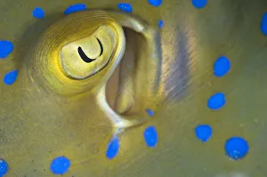
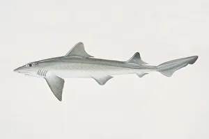
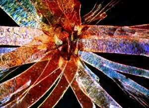
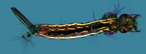
"Discovering the intricate beauty of spiracles in nature's creatures - from the mesmerizing eye and spiracle detail of a Bluespotted Stingray to the sleek side view of a Smooth Dogfish. Delve into the world of tiny organisms like fruit flies and ants, as their delicate they are captured under SEM. Witness the transformation of mosquito larvae, with Anopheles stephensi and Culex showcasing their unique spiracles. Even butterfly larvae like the Large White and December Moth reveal their breathing pores in stunning close-ups. And let's not forget about Silk Moth caterpillars, whose breathing pores become an enchanting focal point. These snapshots remind us that even in small details lies extraordinary wonder. "