
Rod Cell Collection
"Exploring the Intricate World of Rod Cells: A Glimpse into the Retina" In this captivating series of images, we delve into the fascinating realm of rod cells


All Professionally Made to Order for Quick Shipping
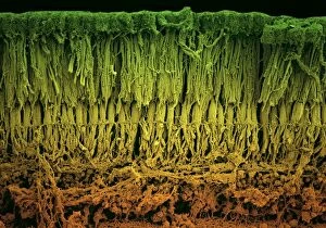
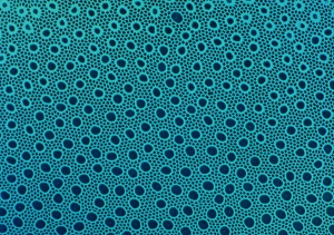
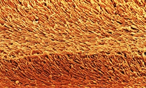
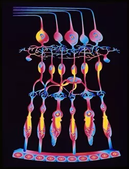
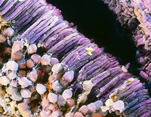
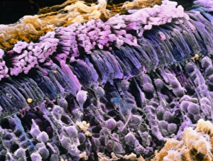
"Exploring the Intricate World of Rod Cells: A Glimpse into the Retina" In this captivating series of images, we delve into the fascinating realm of rod cells, one of the key players in our visual perception. Through scanning electron microscopy (SEM) and transmission electron microscopy (TEM), we uncover intricate details that shed light on their structure and function within the retina. Starting with SEM images C018/0521 and C018/0520, we witness a close-up view of both rod and cone cells residing in different layers of the retina. These delicate structures are responsible for capturing light stimuli and transmitting them to our brain for interpretation. Moving on to TEM images C013/4805 and C013/4804, we zoom in further to observe retinal rod cells at an even higher resolution. Their elongated shape becomes apparent as they stretch across multiple layers within the retina, showcasing their remarkable ability to detect dim light conditions. Next up, we explore developing pig eyes through vibrant light micrographs captured repeatedly. These snapshots reveal how rod cells gradually form during eye development, highlighting nature's meticulous process in creating these essential components for vision. As our journey continues through various microscopic techniques, a high-power light micrograph unveils a mesmerizing glimpse into a monkey's retina. The intricate network formed by countless rod cells is truly awe-inspiring as it demonstrates their abundance within this vital sensory organ. Lastly, colored SEM images provide an artistic perspective on rod cell arrangements within the eye's retina. Vibrant hues accentuate these specialized photoreceptor cells' presence while emphasizing their crucial role in enabling us to see clearly under different lighting conditions. Through these captivating visuals spanning from SEMs to TEMs and colorful representations alike, we gain deeper insight into the complex world of rod cells inhabiting our retinas.