mail_outline sales@mediastorehouse.com
Framed Print
Antique Framed Print
Premium Framed Print
Canvas Print
Metal Print
Photographic Print
Poster Print
Fine Art Print
Mounted Print
Glass Frame
Acrylic Blox
Jigsaw Puzzle
Tote Bag
Photo Mug
Greetings Card
Postcard
Cushion
Mouse Mat
Glass Place Mat
Glass Coaster
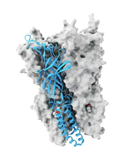
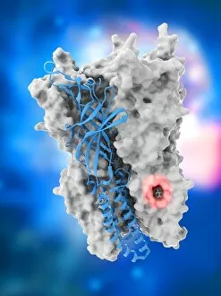
Anaesthetic inhibiting an ion channel C015 / 6718Anaesthetic inhibiting an ion channel. Computer model showing the structure of propofol anaesthetic drug molecules (spheres)
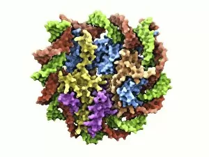
DNA nucleosome, molecular modelDNA nucleosome. Molecular model of a nucleosome, the fundamental repeating unit used to package DNA (deoxyribonucleic acid) inside cell nuclei
DNA nucleosome, molecular model F007 / 9883DNA nucleosome. Molecular model of a nucleosome, the fundamental repeating unit used to package DNA (deoxyribonucleic acid) inside cell nuclei
DNA nucleosome, molecular model F007 / 9888DNA nucleosome. Molecular model of a nucleosome, the fundamental repeating unit used to package DNA (deoxyribonucleic acid) inside cell nuclei
DNA nucleosome, molecular model C016 / 8549DNA nucleosome. Molecular model of a nucleosome, the fundamental repeating unit used to package DNA (deoxyribonucleic acid) inside cell nuclei
Anaesthetic inhibiting an ion channel C015 / 6723Anaesthetic inhibiting an ion channel. Computer model showing the structure of propofol anaesthetic drug molecules (spheres) bound to a pentameric ligand-gated ion channel (pLGIC, blue ribbons)
Anaesthetic inhibiting an ion channel C015 / 6722Anaesthetic inhibiting an ion channel. Computer model showing the structure of propofol anaesthetic drug molecules (spheres) bound to a pentameric ligand-gated ion channel (pLGIC, blue ribbons)
Anaesthetic inhibiting an ion channel C015 / 6720Anaesthetic inhibiting an ion channel. Computer model showing the structure of propofol anaesthetic drug molecules (lower left and right) bound to a pentameric ligand-gated ion channel (pLGIC, grey)
Chromatin fibre, artworkChromatin fibre. Computer artwork of strands of DNA (deoxyribonucleic acid, green) coiled around histone cores (multicoloured) to form a chromatin fibre
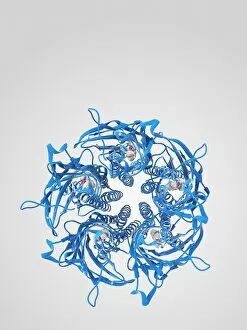
Mouse chromatin protein, molecular modelMouse chromatin protein. Molecular model of the structure of chromatin proteins found in mice. This is similar, but not identical, to the same proteins found in humans
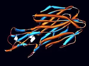
Panton-Valentine toxin. Computer artwork of the ribbon structure of a sub-unit of the Panton- Valentine leucocidin (PVL) toxin from the bacteria Staphylococcus aureus