Fraud Blocker Skip to main content

Respiratory Collection (page 6)

"The Marvels of the Respiratory System: Exploring the Gateway to Life" Diagram of the Lungs and Bronchial Tubes

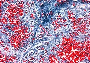
Background imageRespiratory Collection: Bleeding in emphysema

Bleeding in emphysema
Internal bleeding in lung tissue due to pulmonary emphysema, light micrograph. The bleeding is seen as numerous red blood cells (red)

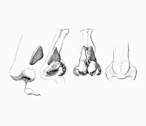
Background imageRespiratory Collection: Noses

Noses, artwork

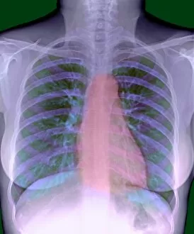
Background imageRespiratory Collection: Heart, chest X-ray

Heart, chest X-ray

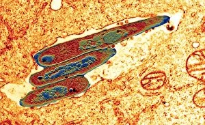
Background imageRespiratory Collection: Macrophage cell engulfing bacteria, TEM

Macrophage cell engulfing bacteria, TEM
Macrophage cell engulfing bacteria. Coloured transmission electron micrograph (TEM) of bacteria (centre, rod-shaped) inside a macrophage cell

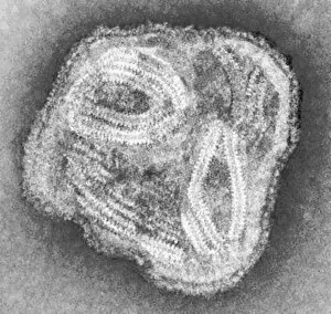
Background imageRespiratory Collection: Paramyxovirus particle, TEM

Paramyxovirus particle, TEM
Paramyxovirus. Transmission electron micrograph (TEM) of a paramyxovirus particle. The internal structure of the virus has been revealed

Background imageRespiratory Collection: Bacterial pneumonia infection

Bacterial pneumonia infection. Light micrograph of pneumonia bacteria (Klebsiella pneumoniae, rod- shaped, pink) among cells in a sample of pus from a pneumonia patient. K

Background imageRespiratory Collection: Deep vein thrombosis, artwork

Deep vein thrombosis, artwork
Deep vein thrombosis. Computer artwork showing blood clots in a leg (brown lumps, bottom right), caused by deep vein thrombosis (DVT), moving to blood vessels in the lungs (blue, top right)

Background imageRespiratory Collection: Avian flu containment, conceptual art

Avian flu containment, conceptual art
Avian flu control, conceptual computer artwork. Quarantine and culling of infected birds can help to slow or prevent the spread of the disease

Background imageRespiratory Collection: Avian flu, conceptual art

Avian flu, conceptual art
Avian flu. Conceptual computer artwork depicting a man in a biohazard uniform walking in front of a giant egg. Avian flu is a viral disease of poultry

Background imageRespiratory Collection: Global avian flu pandemic

Global avian flu pandemic
Global avian flu epidemic, conceptual computer artwork. The H5N1 strain of the avian influenza virus first appeared in humans in Southeast Asia in 1997

Background imageRespiratory Collection: Avian flu

Avian flu. Conceptual computer artwork depicting a giant rooster standing on top of the Earth. Avian flu is a viral disease of poultry

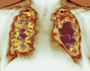
Background imageRespiratory Collection: Loss of lung tissue, X-ray

Loss of lung tissue, X-ray
Lung cavities. Coloured chest X-ray showing large cavities (dark red) in the lungs of a patient in a case of Wegeners granulomatosis

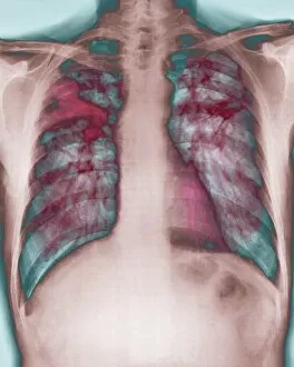
Background imageRespiratory Collection: Fibrosis of the lung, X-ray

Fibrosis of the lung, X-ray
Lung fibrosis. Coloured X-ray of the chest of a patient with pulmonary (lung) fibrosis, or interstitial lung disease. Pulmonary fibrosis is the overgrowth of fibrous scar tissue (dark red)

Background imageRespiratory Collection: AIDS pneumonia infection, TEM

AIDS pneumonia infection, TEM
AIDS pneumonia infection. Coloured transmission electron micrograph (TEM) of a section through Pneumocystis carinii protozoa causing pneumonia. The protozoa are crescent-shaped (red/yellow)

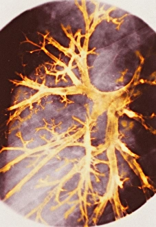
Background imageRespiratory Collection: Bronchitis, X-ray

Bronchitis, X-ray
Bronchitis. Coloured bronchography (X-ray) of swollen bronchioles (orange) in the right lung. Bronchitis causes the network of airways in the lungs to become swollen and produce mucus

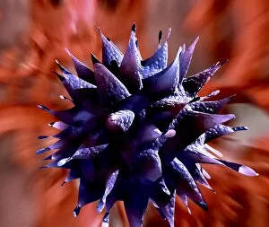
Background imageRespiratory Collection: Influenza virus protein spikes

Influenza virus protein spikes. Computer artwork of part of the surface of a virus particle of the influenza (flu) virus. These spikes (formed by the surface proteins)

Background imageRespiratory Collection: Rhinovirus particles

Rhinovirus particles, computer artwork. This virus consists of a protein capsid (multicoloured), enclosing an RNA (ribonucleic acid) genome

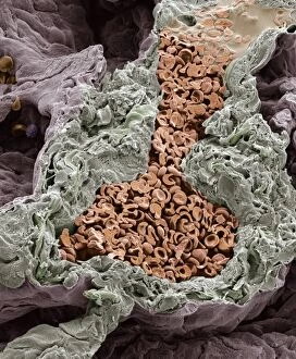
Background imageRespiratory Collection: Blood vessel in the lung, SEM

Blood vessel in the lung, SEM
Blood vessel in the lung, coloured scanning electron micrograph (SEM). This small artery has been fractured to show the enclosed red blood cells (RBCs, orange)

Background imageRespiratory Collection: PM-10 pollutant, artwork

PM-10 pollutant, artwork
PM-10 pollutant. Computer artwork of a microscopic solid pollutant particle. This particle is less than 10 micrometres (a hundredth of a millimetre) in diameter

Background imageRespiratory Collection: Respiratory virus infection treatment

Respiratory virus infection treatment. Computer artwork showing virus particles (spherical) inside a persons nose and how they might be treated by inhaling a drug in the form of a nasal spray

Background imageRespiratory Collection: Tuberculosis diagnosis, 19th century

Tuberculosis diagnosis, 19th century. Artwork showing the French physician Rene Laennec (1781-1826), at the Necker Hospital, Paris, France, examining a patient (left)

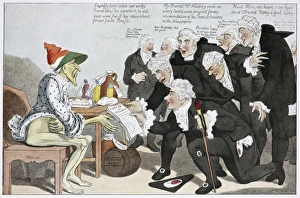
Background imageRespiratory Collection: Influenza epidemic, satirical artwork

Influenza epidemic, satirical artwork
Influenza epidemic. Satirical artwork of the influenza epidemic of 1803, showing a group of British physicians presenting an address of thanks to a figure representing the disease

Background imageRespiratory Collection: Adenovirus particle, TEM

Adenovirus particle, TEM
Adenovirus particle, coloured transmission electron micrograph (TEM). Each particle consists of an icosahedral protein coat (capsid, orange) surrounding double stranded deoxyribonucleic acid (DNA)

Background imageRespiratory Collection: Heart and lungs, historical illustration

Heart and lungs, historical illustration. 19th Century hand coloured lithographic print showing the lungs (left and right) situated either side of the heart (lower centre)

Background imageRespiratory Collection: Chest anatomy, 19th Century illustration

Chest anatomy, 19th Century illustration. Historical hand coloured lithographic print showing the lungs (centre) in the chest, larynx (voicebox, white, upper centre) in the throat

Background imageRespiratory Collection: Respiratory anatomy, 19th Century artwork

Respiratory anatomy, 19th Century artwork. Historical hand coloured lithographic print showing the trachea (wind pipe, vertical) running down from the larynx (voicebox)

Background imageRespiratory Collection: Emphysema, 3D CT scan

Emphysema, 3D CT scan

Background imageRespiratory Collection: Respiratory nerves, 1844 artwork

Respiratory nerves, 1844 artwork
Respiratory nerves. These anatomical artworks are figures 2-3, plate 49, volume 3 (1844) of Traite complet de l anatomie de l homme (1831-1854)

Background imageRespiratory Collection: Lung blood vessel nerves, 1844 artwork

Lung blood vessel nerves, 1844 artwork
Lung blood vessel nerves. This anatomical artwork is figure 4, plate 96 from volume 3 (1844) of Traite complet de l anatomie de l homme (1831-1854)

Background imageRespiratory Collection: H1N1 swine flu virus, SEM

H1N1 swine flu virus, SEM
H1N1 swine flu virus. Coloured scanning electron micrograph (SEM) of swine influenza (flu) virus particles (virions, yellow)

Background imageRespiratory Collection: Climbing perch

Climbing perch. These fish, from the genus Anabas, are found in southern Asia, including India, Burma, Indonesia and the Philippines. The specimens shown here are from India

Background imageRespiratory Collection: Adenovirus particles, TEM

Adenovirus particles, TEM
Adenovirus particles. Coloured transmission electron micrograph (TEM) of adenovirus particles. Each particle consists of an icosahedral protein coat (capsid)

Background imageRespiratory Collection: Legionella bacteria, SEM

Legionella bacteria, SEM
Legionella bacteria. Coloured scanning electron micrograph (SEM) of Legionella pneumophila bacteria, the cause of Legionnaires disease

Background imageRespiratory Collection: La Crosse encephalitis virus, TEM

La Crosse encephalitis virus, TEM
La Crosse encephalitis virus. Coloured transmission electron micrograph (TEM) of La Cross (LAC) encephalitis virus particles (virions)

Background imageRespiratory Collection: Legionella bacteria, light micrograph

Legionella bacteria, light micrograph
Legionella bacteria. Coloured light micrograph of Legionella pneumophila bacteria, the cause of Legionnaires disease. This rod-shaped

Background imageRespiratory Collection: California encephalitis virus, TEM

California encephalitis virus, TEM
California encephalitis virus. Coloured transmission electron micrograph of California encephalitis virus particles (virions)


All Professionally Made to Order for Quick Shipping

"The Marvels of the Respiratory System: Exploring the Gateway to Life" Diagram of the Lungs and Bronchial Tubes: Unveiling the intricate network within our bodies that facilitates every breath we take, this diagram showcases the lungs and bronchial tubes, highlighting their vital role in oxygenating our bloodstream. Diagram of the Heart, Lungs, and Windpipe: Witnessing a symphony of life-sustaining organs working harmoniously together, this diagram depicts the heart pumping blood while being surrounded by the lungs and windpipe – a testament to our body's remarkable respiratory system. Illustration of Human Respiratory System: Embark on an anatomical journey through our respiratory system as this illustration reveals its various components such as oral cavity, nasal cavity, larynx, trachea, bronchus, and lungs – each playing a crucial part in ensuring efficient respiration. Tension Pneumothorax X-ray: Peering into medical imaging technology's window into diagnosis and treatment strategies for collapsed lungs caused by tension pneumothorax - an urgent condition requiring immediate attention to restore normal breathing patterns. English Oak Leaf Pores SEM Image: Delving into nature's wonders at microscopic levels reveals mesmerizing images showcasing English oak leaf pores under scanning electron microscopy (SEM), reminding us how even plants have their unique respiratory systems. Cystic Fibrosis: Unmasking a Genetic Challenge Exploring cystic fibrosis - a genetic disorder affecting lung function - sheds light on ongoing research efforts aimed at improving treatments for individuals facing challenges with their respiratory health from an early age. Konstantin Buteyko: Soviet Doctor Revolutionizing Breathing Techniques Discovering how Dr. Konstantin Buteyko revolutionized breathing techniques offers insights into his innovative methods that have helped countless individuals manage asthma symptoms and enhance overall respiratory well-being. Paramyxovirus Particles TEM Image.