Fraud Blocker Skip to main content

Radiology Collection (page 6)

Radiology, the art of peering into the human body's intricate mysteries, reveals a world unseen

Background imageRadiology Collection: Taking an X-ray

Taking an X-ray
MODEL RELEASED. Taking an X-ray. Radiographer (standing) taking an X-ray of a female patient. The device held by the radiographer passes X-rays through the patients body

Background imageRadiology Collection: Pulmonary consolidation, X-ray

Pulmonary consolidation, X-ray
Pulmonary consolidation. X-ray showing that the upper lobe of a patients left lung has become consolidated (filled with pus) due to pneumonia

Background imageRadiology Collection: Childhood rickets, X-ray

Childhood rickets, X-ray
Childhood rickets. Coloured frontal X-ray of the legs of a 3-year-old child with rickets. Rickets (Osteomalacia) occurs predominantly in children

Background imageRadiology Collection: Arteritis of the knee, X-ray

Arteritis of the knee, X-ray
Arteritis of the knee. Coloured X-ray image of the knee of an 85-year-old patient with arteritis. Arteritis is an inflammation of the walls of arteries

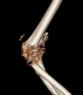
Background imageRadiology Collection: Fractured elbow, CT scan

Fractured elbow, CT scan
Fractured elbow. Coloured 3D computed tomography (CT) scan of the elbow of an 84-year-old patient that was fractured after a fall

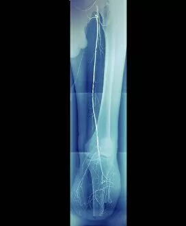
Background imageRadiology Collection: Diabetic amputee, angiogram

Diabetic amputee, angiogram
Diabetic amputee. Coloured angiogram X-ray of the left leg of a 54-year-old diabetic smoker and alcoholic who has had their leg amputated below the knee (lower centre)

Background imageRadiology Collection: Takayasus arteritis, 3d CT scan

Takayasus arteritis, 3d CT scan
Takayasus arteritis. Coloured 3d computed tomography (CT) angiogram of the arteries of the neck and head of a 56-year-old patient with Takayasus arteritis

Background imageRadiology Collection: Tourettes treatment, X-ray

Tourettes treatment, X-ray
Tourettes treatment, coloured X-ray. Profile view of the skull of a 25-year-old man with Tourettes syndrome, showing electrodes implanted in his brain

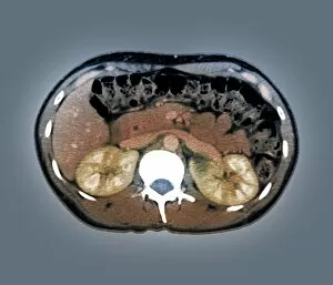
Background imageRadiology Collection: Medullary sponge kidney, CT scan

Medullary sponge kidney, CT scan
Medullary sponge kidney. Coloured computed tomography (CT) scan in axial section of the abdomen of a 25-year-old patient with medullary sponge kidney, or Cacchi Ricci disease

Background imageRadiology Collection: Chronic liver disease, CT scan

Chronic liver disease, CT scan
Chronic liver disease. Coloured computed tomography (CT) scan of an axial section through the abdominopelvic region of a 58-year-old patient with a history of hepatitis C

Background imageRadiology Collection: Slipped disc, MRI scan

Slipped disc, MRI scan
Slipped disc, coloured magnetic resonance imaging (MRI) scan. Profile view of the spine of a 59-year-old patient with severe rheumatoid arthritis and a herniated (slipped) disc (red)

Background imageRadiology Collection: Healthy liver, CT scan

Healthy liver, CT scan
Healthy live. Coloured computed tomography (CT) scan of section through the abdomen of a healthy 42-year-old, showing their liver (upper left and centre)

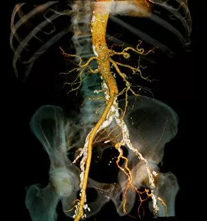
Background imageRadiology Collection: Arteritis, 3d CT scan

Arteritis, 3d CT scan
Arteritis. Coloured 3d computed tomography (CT) angiogram of the abdominal aorta in a 60-year-old patient after treatment for arteritis

Background imageRadiology Collection: Aortic aneurysm CT scan

Aortic aneurysm CT scan
Aortic aneurysm, coloured 3D computed tomography (CT) scan. Side view of the abdomen of a 65-year-old patient with an extended aneurysm of the aorta

Background imageRadiology Collection: Charcot arthropathy, MRI scans

Charcot arthropathy, MRI scans
Charcot arthropathy. Coloured magnetic resonance imaging (MRI) scans of the foot of a 33-year-old patient with Charcot arthropathy

Background imageRadiology Collection: Spine in Hurler Syndrome, X-ray

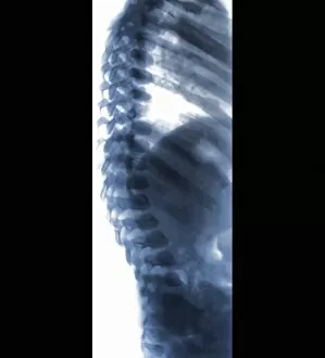
Spine in Hurler Syndrome, X-ray
Spine in Hurler Syndrome. Coloured X-ray of the spine of a 26-month-old child with Hurler syndrome (also called mucopolysaccharidosis type I, MPS I)

Background imageRadiology Collection: Fractured femur, X-ray

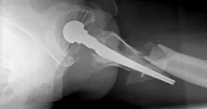
Fractured femur, X-ray
Fractured femur. X-ray of a patient with a total hip replacement who has fractured their femur (thigh bone). The fracture (centre right) has occurred towards the end of the implant (solid white)

Background imageRadiology Collection: Dislocated hip replacement, X-ray

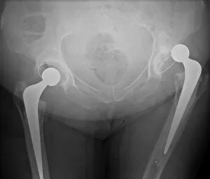
Dislocated hip replacement, X-ray
Dislocated hip replacement. Frontal X-ray of the pelvis of a patient with bi-lateral (both sides) total hip replacements. The right joint replacement (right) has become dislocated

Background imageRadiology Collection: 3D hospital scan, conceptual artwork

3D hospital scan, conceptual artwork. Computer artwork of a 3-dimensional image of a human, showing both the muscle and skeletal structure, with X-ray artworks above

Background imageRadiology Collection: Examining X-ray

Examining X-ray
MODEL RELEASED. Examining X-ray. Radiologist examining an X-ray of a patients hand and wrist, looking for broken bones and other disorders


All Professionally Made to Order for Quick Shipping

Radiology, the art of peering into the human body's intricate mysteries, reveals a world unseen. From panoramic dental X-rays capturing every tooth's tale to an assortment of coloured X-rays and body scans painting a vibrant picture of our inner workings, this field never ceases to amaze. Intriguingly, even mundane ailments like headaches can transform into captivating X-ray artwork that showcases the hidden source of pain. Hands gracefully suspended in time through an X-ray lens remind us of their tireless work in our daily lives. A healthy knee caught on film reminds us of the marvels within our own bodies. Conversely, tension pneumothorax exposes a fragile balance disrupted by life-threatening conditions that require immediate attention. Picture No. 11676149 captures a moment frozen in time—a snapshot revealing secrets only visible through radiology's watchful eye. Bacterial meningitis unravels its sinister plot under the scrutiny of an MRI scan while total knee replacement unveils newfound mobility through meticulous X-rays. Delving deeper into history, we find ourselves transported to 1900 when soldiers with bullet-bound wounds were examined using early forms of X-ray technology—an extraordinary testament to radiology's role in saving lives during times of conflict. The elegance continues as we witness a healthy ankle joint basking in its uninhibited movement—a reminder that sometimes it is what lies beneath that truly matters. And scoliosis presents itself boldly on an X-ray canvas—each curve telling a unique story etched upon the spine's landscape. Radiology holds within it countless tales waiting to be discovered—one image at a time, and is both science and art intertwined—a window into humanity's most intimate details and profound struggles. With each click and flash, radiologists illuminate paths towards healing and understanding—the guardians who unlock the secrets concealed within our very bones.