Fraud Blocker Skip to main content

Radiology Collection (page 3)

Radiology, the art of peering into the human body's intricate mysteries, reveals a world unseen

Background imageRadiology Collection: Stroke, CT scans

Stroke, CT scans
Stroke. Coloured computed tomography (CT) scans of sections through the brain of a patient taken 24 hours, 10 days, 25 days, and 90 days after a haemorrhagic stroke

Background imageRadiology Collection: Coronary stenosis treatment, X-rays

Coronary stenosis treatment, X-rays
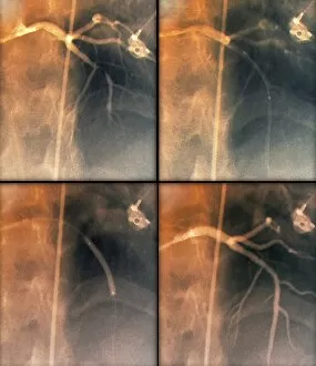
Coronary stenosis treatment. Coloured angiograms showing the left coronary artery in the heart of a 60-year-old patient before (top left)

Background imageRadiology Collection: Fibrolipoma of the spine, MRI scan

Fibrolipoma of the spine, MRI scan
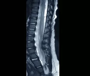
Fibrolipoma of the spine. Coloured magnetic resonance imaging (MRI) scan of a sagittal section through the thoraco-lumbar spine of a 19-month-old patient

Background imageRadiology Collection: Total knee replacement, X-ray

Total knee replacement, X-ray
Total knee replacement. X-ray of the legs of a 49-year-old patient who has had a total knee replacement (white) of the left knee (right)

Background imageRadiology Collection: Mammogram, conceptual image

Mammogram, conceptual image. Computer artwork of a womans right breast in an X-ray machine during a mammogram (breast X-ray)

Background imageRadiology Collection: Diagnostics imaging techniques, concept C016 / 7511

Diagnostics imaging techniques, concept C016 / 7511
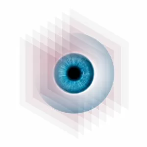
Diagnostics imaging techniques, conceptual computer artwork

Background imageRadiology Collection: Colon cancer, barium contrast CT scan

Colon cancer, barium contrast CT scan
Colon cancer. Coloured barium contrast computed tomography (CT) scan, and 3D CT colonoscopy image (inset), of an axial section through the sigmoid colon (large intestine) of a 54-year-old patient

Background imageRadiology Collection: Colon cancer, 3D colonoscopy image

Colon cancer, 3D colonoscopy image
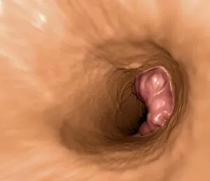
Colon cancer. 3D colonoscopy image showing the location of a cancerous growth (centre right) in the colon of a 59-year-old patient. Here, the growth has caused a narrowing (stenosis) of the colon

Background imageRadiology Collection: Total hip replacement, X-rays

Total hip replacement, X-rays
Total hip replacement. Coloured X-rays taken during (left) and after (right) surgery to replace the right hip joint of a 65-year-old patient using a prosthetic implant

Background imageRadiology Collection: Hearing aid, X-ray

Hearing aid, X-ray
Hearing aid. Coloured X-ray of the head of a 70-year-old patient, showing a hearing aid (white, centre left) located behind their left ear

Background imageRadiology Collection: Breast lumps, MRI

Breast lumps, MRI
Breast lumps. Coloured magnetic resonance imaging (MRI) scan of the chest of a 20-year-old patient, showing multiple fibroadenomas (light patches) in both breasts

Background imageRadiology Collection: Narrowed aorta, 3D CT scans

Narrowed aorta, 3D CT scans
Narrowed aorta. Coloured 3D computed tomography (CT) scans of the heart (centre of each scan) and major blood vessels of a 50-year-old patient

Background imageRadiology Collection: Targeted breast surgery, mammogram

Targeted breast surgery, mammogram
Targeted breast surgery. Coloured mammograms of the right breast of a 47-year-old female patient being used to locate the exact site of micro-lesions, prior to surgery

Background imageRadiology Collection: Colon polyp, 3D colonoscopy image

Colon polyp, 3D colonoscopy image
Colon polyp. Coloured 3D computed tomography (CT) colonoscopy image of a pedunculated (stalked) polyp in the sigmoid colon (large intestine) of a 50-year-old patient

Background imageRadiology Collection: Failed hip replacement, CT scan

Failed hip replacement, CT scan
Failed hip replacement. Coloured computed tomography (CT) scan of a total hip replacement (solid white) in a 70-year-old patient

Background imageRadiology Collection: Calcified heart valve, CT scan

Calcified heart valve, CT scan
Calcified heart valve. Coloured computed tomography (CT) scan of a section through the heart of a 67-year-old patient showing calcification of the aortic valve (round)

Background imageRadiology Collection: Brain tumour, MRI scans

Brain tumour, MRI scans
Brain tumour. Coloured magnetic resonance imaging (MRI) scans of sections through the head of a 23-year-old patient with a lesion (dark patches)

Background imageRadiology Collection: Stroke, MRI scan

Stroke, MRI scan
Stroke. Coloured magnetic resonance imaging (MRI, left) and computed tomography (CT, right) scans of axial sections through the brain of a 58-year-old patient

Background imageRadiology Collection: Berry aneurysm, MRI scan

Berry aneurysm, MRI scan
Stroke. Coloured computed tomography (CT) scan of an axial section through the brain of a 58-year-old patient, showing the damage (dark) caused 24 hours after a cerebral infarction

Background imageRadiology Collection: Creutzfeldt-Jakob disease, MRI scan

Creutzfeldt-Jakob disease, MRI scan
Creutzfeldt-Jakob disease. Coloured magnetic resonance imaging (MRI) scan of an axial section through the brain of a 35-year-old patient

Background imageRadiology Collection: Stroke, CT scan

Stroke, CT scan
Stroke. Coloured computed tomography (CT) scan of an axial section through the brain of a 58-year-old patient, showing the damage (purple) caused 24 hours after a cerebral infarction

Background imageRadiology Collection: Double mastectomy breast implants, MRI

Double mastectomy breast implants, MRI
Double mastectomy breast implants. Post-operative coloured magnetic resonance imaging (MRI) scan of a section through the chest of a 61-year-old patient after a double mastectomy (breast removal)

Background imageRadiology Collection: Kidney tumour, CT scan

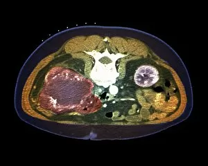
Kidney tumour, CT scan
Kidney tumour. Coloured computed tomography (CT) scan of a section through the abdomen of a 62-year-old patient, showing necrosis (death) of tissue in the right kidney

Background imageRadiology Collection: Hip fracture reduction, 3D CT scan

Hip fracture reduction, 3D CT scan
Hip fracture reduction. Coloured 3D computed tomography (CT) scan of the pelvis of an 81-year-old patient with a fracture to the neck of the right femur (thigh bone)

Background imageRadiology Collection: Secondary lung cancer, X-ray

Secondary lung cancer, X-ray
Secondary lung cancers. Coloured X-ray of malignant (cancerous) tumours (red patches) in the lungs of a 60-year-old female patient

Background imageRadiology Collection: Calcified heart valve, CT scans

Calcified heart valve, CT scans
Calcified heart valve. Coloured computed tomography (CT) scans of a section through the heart of a 67-year-old patient showing calcification of the aortic valve (round)

Background imageRadiology Collection: Colon cancer, CT scans

Colon cancer, CT scans
Colon cancer. 2D computed tomography (CT) rendering (left) and 3D colonoscopy image (right) showing the location of a cancerous growth in the colon of a 59-year-old patient

Background imageRadiology Collection: Uterine fibroid, X-ray C014 / 4919

Uterine fibroid, X-ray C014 / 4919
Uterus tumour. Coloured X-ray of the abdomen of a woman with a fibroid (round, centre) in her uterus. A fibroid is a fibrous benign tumour originating from muscular tissue (leimyoma)

Background imageRadiology Collection: Uterine fibroid, X-ray C014 / 4918

Uterine fibroid, X-ray C014 / 4918
Uterus tumour. Coloured X-ray of the abdomen of a woman with a fibroid (orange, round) in her uterus. A fibroid is a fibrous benign tumour originating from muscular tissue (leimyoma)

Background imageRadiology Collection: Endometriosis, X-ray C014 / 4917

Endometriosis, X-ray C014 / 4917
Endometriosis. Coloured X-ray of the abdomen of woman with endometriosis. Endometriosis is the movement of cells from the lining of the uterus to the abdominal cavity

Background imageRadiology Collection: Endometriosis, X-ray C014 / 4916

Endometriosis, X-ray C014 / 4916
Endometriosis. Coloured X-ray of the abdomen of woman with endometriosis. Endometriosis is the movement of cells from the lining of the uterus to the abdominal cavity

Background imageRadiology Collection: Stomach cancer, barium X-ray

Stomach cancer, barium X-ray
Stomach cancer. Coloured frontal X-ray of the upper gastroduodenal tract of a patient with stomach cancer (grey, upper centre)

Background imageRadiology Collection: Radiotherapy, conceptual image C013 / 5587

Radiotherapy, conceptual image C013 / 5587
Radiotherapy, conceptual image. Computer artwork representing the use of ionising radiation to kill cancerous cells (centre)

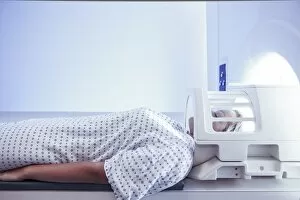
Background imageRadiology Collection: MRI scan

MRI scan

Background imageRadiology Collection: IPhone 4, coloured x-ray C016 / 7214

IPhone 4, coloured x-ray C016 / 7214
Coloured x-ray of an iPhone 4, showing the internal structure. The iPhone 4 mobile phone was introduced on 24 June 2010. It is a smart phone

Background imageRadiology Collection: IPhone, X-ray C016 / 7207

IPhone, X-ray C016 / 7207
X-ray of an iPhone 4, showing the internal structure. The iPhone 4 mobile phone was introduced on 24 June 2010. It is a smart phone

Background imageRadiology Collection: Dental X-rays

Dental X-rays

Background imageRadiology Collection: Uterine bleeding treatment, DSA scan

Uterine bleeding treatment, DSA scan
Uterine bleeding treatment. Coloured digital subtraction angiogram (DSA) of the pelvic region of a 45-year-old woman with menometrorrhagia, showing embolisation of the uterine arteries

Background imageRadiology Collection: Ionisation chamber system C016 / 3789

Ionisation chamber system C016 / 3789
Ionisation chamber system (white, left) and viewing the resulting data on a screen (right). Ionisation chambers enable accurate measurement of the amount of radiation absorbed by an object or organic

Background imageRadiology Collection: Ionisation chamber system C016 / 3792

Ionisation chamber system C016 / 3792
Ionisation chamber system. Robotic arm of an automatic sample changer loading a radioactive sample into a secondary standard ionisation chamber

Background imageRadiology Collection: Roosevelt bullet, 1912 chest X-ray C016 / 2549

Roosevelt bullet, 1912 chest X-ray C016 / 2549
Roosevelt bullet. Chest X-ray showing the bullet used in the non-fatal shooting of former US President Theodore Roosevelt (1858-1919)

Background imageRadiology Collection: Mobile X-ray unit, World War I C016 / 2548

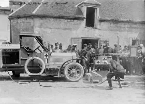
Mobile X-ray unit, World War I C016 / 2548
Mobile X-ray unit. French soldiers assembled around a mobile X-ray unit during World War I (1914-1918). X-rays had been discovered in 1895 by German physicist Wilhelm Conrad Roentgen

Background imageRadiology Collection: Doctor examining an X-ray C013 / 8846

Doctor examining an X-ray C013 / 8846
Doctor examining an X-ray

Background imageRadiology Collection: Doctor examining an X-ray C013 / 8845

Doctor examining an X-ray C013 / 8845
Doctor examining an X-ray

Background imageRadiology Collection: Hip replacement surgery C013 / 4962

Hip replacement surgery C013 / 4962
Hip replacement surgery. Surgeon looking at an X-ray of the hip of a patient undergoing hip replacement surgery

Background imageRadiology Collection: Womans legs, with knee X-ray

Womans legs, with knee X-ray
MODEL RELEASED. Womans legs superimposed on an X-ray of a knee joint. X-rays can be used to diagnose disorders of the skeletal system

Background imageRadiology Collection: Adult and child hand X-rays

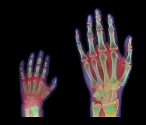
Adult and child hand X-rays
Adult and child hands. Coloured X-rays of healthy human hands at 3 years (left) and at 20 years. Bones are green with flesh red


All Professionally Made to Order for Quick Shipping

Radiology, the art of peering into the human body's intricate mysteries, reveals a world unseen. From panoramic dental X-rays capturing every tooth's tale to an assortment of coloured X-rays and body scans painting a vibrant picture of our inner workings, this field never ceases to amaze. Intriguingly, even mundane ailments like headaches can transform into captivating X-ray artwork that showcases the hidden source of pain. Hands gracefully suspended in time through an X-ray lens remind us of their tireless work in our daily lives. A healthy knee caught on film reminds us of the marvels within our own bodies. Conversely, tension pneumothorax exposes a fragile balance disrupted by life-threatening conditions that require immediate attention. Picture No. 11676149 captures a moment frozen in time—a snapshot revealing secrets only visible through radiology's watchful eye. Bacterial meningitis unravels its sinister plot under the scrutiny of an MRI scan while total knee replacement unveils newfound mobility through meticulous X-rays. Delving deeper into history, we find ourselves transported to 1900 when soldiers with bullet-bound wounds were examined using early forms of X-ray technology—an extraordinary testament to radiology's role in saving lives during times of conflict. The elegance continues as we witness a healthy ankle joint basking in its uninhibited movement—a reminder that sometimes it is what lies beneath that truly matters. And scoliosis presents itself boldly on an X-ray canvas—each curve telling a unique story etched upon the spine's landscape. Radiology holds within it countless tales waiting to be discovered—one image at a time, and is both science and art intertwined—a window into humanity's most intimate details and profound struggles. With each click and flash, radiologists illuminate paths towards healing and understanding—the guardians who unlock the secrets concealed within our very bones.