Fraud Blocker Skip to main content

Platelets Collection

Platelets play a crucial role in the intricate process of blood coagulation cascade

Background imagePlatelets Collection: Blood clot, SEM

Blood clot, SEM
Blood clot, coloured scanning electron micrograph (SEM). Red blood cells are red and fibrin protein strands are green. Platelets are at bottom right

Background imagePlatelets Collection: Blood coagulation cascade, artwork C016 / 9873

Blood coagulation cascade, artwork C016 / 9873
Blood coagulation cascade. Artwork of the biochemical cascade of blood chemicals and proteins during blood clotting (coagulation). The blood vessel and its layered wall is at upper left

Background imagePlatelets Collection: Blood clot, SEM C016 / 9747

Blood clot, SEM C016 / 9747
Blood clot, coloured scanning electron micrograph (SEM). Red blood cells (erythrocytes) are trapped within a fibrin protein mesh (beige)

Background imagePlatelets Collection: Lung tissue, TEM

Lung tissue, TEM
Lung tissue, coloured transmission electron micrograph (TEM). A single alveolus is at center. At top right is a pulmonary capillary containing a red blood cell (red) and a platelet (yellow)

Background imagePlatelets Collection: White blood cells and platelets, SEM C016 / 3099

White blood cells and platelets, SEM C016 / 3099
White blood cells and platelets. Coloured scanning electron micrograph (SEM) of platelets (green) with a variety of different white blood cells (leucocytes, yellow)

Background imagePlatelets Collection: White blood cells and platelets, SEM C016 / 3098

White blood cells and platelets, SEM C016 / 3098
White blood cells and platelets. Coloured scanning electron micrograph (SEM) of platelets (purple) with a variety of different white blood cells (leucocytes, blue)

Background imagePlatelets Collection: Blood platelets

Blood platelets. Coloured scanning electron micro- graph (SEM) of activated blood platelets. Platelet cells (thrombocytes) are formed in the bone marrow

Background imagePlatelets Collection: Blood clot, SEM C016 / 9751

Blood clot, SEM C016 / 9751
Blood clot, coloured scanning electron micrograph (SEM). Red blood cells (erythrocytes) are trapped within a fibrin protein mesh (beige)

Background imagePlatelets Collection: Blood clot, SEM C016 / 9746

Blood clot, SEM C016 / 9746
Blood clot, coloured scanning electron micrograph (SEM). Red blood cells (erythrocytes) are trapped within a fibrin protein mesh (beige)

Background imagePlatelets Collection: Blood clot, SEM C016 / 9749

Blood clot, SEM C016 / 9749
Blood clot, coloured scanning electron micrograph (SEM). Red blood cells (erythrocytes) are trapped within a fibrin protein mesh (beige)

Background imagePlatelets Collection: Blood clot, SEM C017 / 7141

Blood clot, SEM C017 / 7141
Blood clot, coloured scanning electron micrograph (SEM). Red blood cells (erythrocytes) and a white blood cell (purple) trapped within a fibrin protein mesh (cream)

Background imagePlatelets Collection: Blood clot, SEM C016 / 9750

Blood clot, SEM C016 / 9750
Blood clot, coloured scanning electron micrograph (SEM). Red blood cells (erythrocytes) are trapped within a fibrin protein mesh (beige)

Background imagePlatelets Collection: Blood clot, SEM C016 / 9753

Blood clot, SEM C016 / 9753
Blood clot. Coloured scanning electron micrograph (SEM) of a blood clot from the inner wall of the left ventricle of a heart

Background imagePlatelets Collection: Blood clot, SEM C016 / 9752

Blood clot, SEM C016 / 9752
Blood clot, coloured scanning electron micrograph (SEM). Red blood cells (erythrocytes) are trapped within a fibrin protein mesh (beige)

Background imagePlatelets Collection: Blood clot, SEM C016 / 9745

Blood clot, SEM C016 / 9745
Blood clot, coloured scanning electron micrograph (SEM). Red blood cells (erythrocytes) are trapped within a fibrin protein mesh (beige)

Background imagePlatelets Collection: Blood clot, SEM C016 / 9748

Blood clot, SEM C016 / 9748
Blood clot, coloured scanning electron micrograph (SEM). Red blood cells (erythrocytes) are trapped within a fibrin protein mesh (beige)

Background imagePlatelets Collection: Blood clot, SEM C015 / 9608

Blood clot, SEM C015 / 9608
Blood clot. Coloured scanning electron micrograph (SEM) of a blood clot from the inner wall of the left ventricle of a heart

Background imagePlatelets Collection: Blood cells, illustration C018 / 0802

Blood cells, illustration C018 / 0802
Blood cells. All cellular blood components originate from the same cell, the haematopoietic stem cell. The stem cell differentiates into two types of progenitor cells

Background imagePlatelets Collection: Blood clot, SEM C016 / 9744

Blood clot, SEM C016 / 9744
Blood clot, coloured scanning electron micrograph (SEM). Red blood cells (erythrocytes) are trapped within a fibrin protein mesh (beige)

Background imagePlatelets Collection: White blood cells, SEM C016 / 3102

White blood cells, SEM C016 / 3102
White blood cells. Coloured scanning electron micrograph (SEM) of white blood cells (leucocytes) from a sample of peripheral blood

Background imagePlatelets Collection: Blood cells, SEM C016 / 3101

Blood cells, SEM C016 / 3101
Blood cells. Coloured scanning electron micrograph (SEM) of a variety of different white blood cells (leucocytes, blue/purple) with two red blood cells (erythrocytes)

Background imagePlatelets Collection: Blood cells, SEM C016 / 3100

Blood cells, SEM C016 / 3100
Blood cells. Coloured scanning electron micrograph (SEM) of a variety of different white blood cells (leucocytes, yellow/green) with two red blood cells (erythrocytes)

Background imagePlatelets Collection: Macrophage and platelets, SEM C016 / 3094

Macrophage and platelets, SEM C016 / 3094
Macrophage and platelets. Coloured scanning electron micrograph (SEM) of platelets (green) attached to a macrophage white blood cell (turquoise)

Background imagePlatelets Collection: Platelets, SEM C016 / 3091

Platelets, SEM C016 / 3091
Platelets, coloured scanning electron micrograph (SEM). Platelets, or thrombocytes, are fragments of white blood cells. When blood clots

Background imagePlatelets Collection: Blood clot C013 / 7129

Blood clot C013 / 7129
Blood clot. Coloured scanning electron micrograph (SEM) of a blood clot (centre) forming in connective tissue. Red blood cells (erythrocytes)

Background imagePlatelets Collection: Blood clot C013 / 7128

Blood clot C013 / 7128
Blood clot. Coloured scanning electron micrograph (SEM) of a blood clot (centre) forming in connective tissue. Red blood cells (erythrocytes)

Background imagePlatelets Collection: Blood cells, artwork C013 / 4726

Blood cells, artwork C013 / 4726
Blood cells. Computer artwork of red blood cells (erythrocytes, red), white blood cells (leucocytes, white), and platelets (thrombocytes, yellow)

Background imagePlatelets Collection: Blood cells, artwork C013 / 4707

Blood cells, artwork C013 / 4707
Blood cells. Computer artwork of red blood cells (erythrocytes, red), white blood cells (leucocytes, white), and platelets (thrombocytes, yellow)

Background imagePlatelets Collection: Blood vessel and cells, computer artwork

Blood vessel and cells, computer artwork
Blood vessel and cells. Computer artwork of various blood cells and a blood vessel. The blood vessel consists of an outer layer of muscle (pink)

Background imagePlatelets Collection: F / col TEM Unactivated blood platelets

F / col TEM Unactivated blood platelets
False-colour transmission electron micrograph of unactivated blood platelets & some red blood cells (bottom left). Platelets (thrombocytes) are small

Background imagePlatelets Collection: Blood clots, SEM

Blood clots, SEM
Blood clots. Coloured scanning electron micrograph (SEM) of small blood clots (red) in a blood vessel. The clots consist of red blood cells, white blood cells, and platelets

Background imagePlatelets Collection: Platelets in a blood clot

Platelets in a blood clot. Coloured scanning electron micrograph (SEM) of platelet cells (thrombocytes) in a blood clot. They are oval shaped

Background imagePlatelets Collection: F / col TEM of section through blood platelets

F / col TEM of section through blood platelets
False colour transmission electron micrograph of a section through activated blood platelets. Platelets (thrombocytes) are formed in the bone marrow, and circulate in blood in large numbers

Background imagePlatelets Collection: Sickle cell anaemia

Sickle cell anaemia. Artwork of red blood cells in sickle cell anaemia (drepanocytosis). The sickle- shaped red blood cells (some seen at upper left) contrast with normal red blood cells (rounded)

Background imagePlatelets Collection: Blood platelet, TEM

Blood platelet, TEM

Background imagePlatelets Collection: Coloured SEM of an activated blood platelet

Coloured SEM of an activated blood platelet

Background imagePlatelets Collection: Coloured SEM of activated platelets in human blood

Coloured SEM of activated platelets in human blood

Background imagePlatelets Collection: Blood cells, computer artwork

Blood cells, computer artwork
Blood cells. Computer artwork of red blood cells, white blood, white blood cells (blue) and platelets (yellow) travelling through the lumen of the blood vessel

Background imagePlatelets Collection: Coronary thrombosis, light micrograph

Coronary thrombosis, light micrograph
Coronary thrombosis. Light micrograph of a section through a thrombus (blood clot) in the coronary artery of the heart. The clot is packed full of red blood cells (red)

Background imagePlatelets Collection: Platelets, SEM

Platelets, SEM
Platelets. Coloured scanning electron micrograph (SEM) of a clump of activated platelets from a blood clot. Platelets are fragments of white blood cells

Background imagePlatelets Collection: Activated platelets, SEM

Activated platelets, SEM
Activated platelets. Coloured scanning electron micrograph (SEM) of activated platelets, or thrombocytes, starting to adhere to exposed sub-endothelium on the inner surface of a vein

Background imagePlatelets Collection: Platelet activation, artwork

Platelet activation, artwork
actication of PRP gel

Background imagePlatelets Collection: Blood clotting, SEM

Blood clotting, SEM
Blood clotting. Coloured scanning electron micrograph (SEM) of human red blood cells (erythrocytes, red) and platelets (thrombocyte, white) forming a blood clot

Background imagePlatelets Collection: Red blood cells and platelets, SEM

Red blood cells and platelets, SEM
Red blood cells and platelets. Coloured scanning electon micrograph (SEM) of human erythrocytes (red blood cells) and a platelet aggregate (purple)

Background imagePlatelets Collection: Activated platelets, artwork

Activated platelets, artwork
Activated platelets. Artwork of platelets (thrombocytes) that have been activated. Platelets are part of the blood. When a blood vessel is damaged

Background imagePlatelets Collection: Blood cells in blood vessel, artwork

Blood cells in blood vessel, artwork
Blood cells in blood vessel, computer artwork. Red blood cells (erythrocytes, red) distribute oxygen to body tissues, and carry waste carbon dioxide back to the lungs


All Professionally Made to Order for Quick Shipping

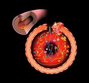
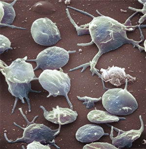
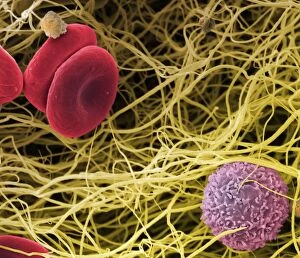
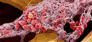
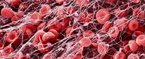
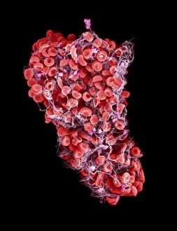
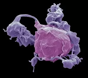
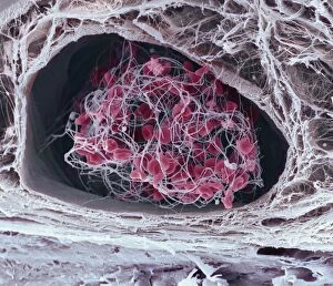
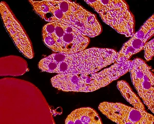
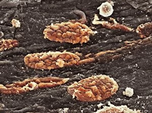
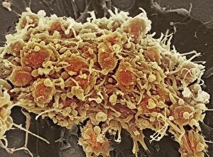
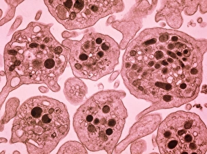
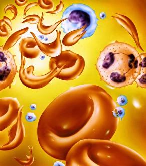
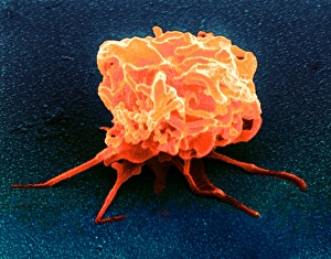
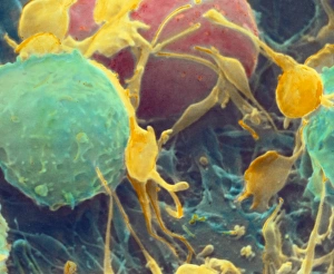
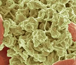
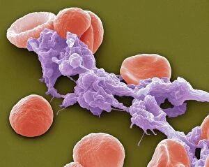
Platelets play a crucial role in the intricate process of blood coagulation cascade. These tiny, disc-shaped cells are like the unsung heroes of our circulatory system, working tirelessly to prevent excessive bleeding and promote healing. In the mesmerizing artwork C016 / 9873, platelets take center stage as they gather around a blood clot, their mission clear: to seal off any wounds and restore balance within our bodies. This microscopic view captured by SEM reveals their intricate structure and highlights their importance in maintaining our health. When an injury occurs, platelets rush to the scene, forming a plug that stops bleeding. In SEM images such as C016 / 9747 or C017 / 7141, we witness these remarkable cells interweaving with fibrin strands to create a robust blood clot, and is through this collaboration between platelets and other components of our blood that we can avoid potentially life-threatening hemorrhages. SEM images like C016 / 9750 or C016 / 9752 showcase platelets alongside white blood cells – another essential component of our immune system. Together, they form an army against infections and work synergistically to maintain homeostasis within us. As we marvel at these captivating visuals provided by science, let us not forget the vital role played by platelets in safeguarding our well-being. They may be small individually but collectively possess immense power when it comes to ensuring proper wound healing and preventing excessive bleeding. So next time you see a drop of your own precious red fluid escaping from your body's protective barrier, remember those incredible little warriors called platelets who come rushing in for your rescue.