
Phospholipid Collection
Phospholipids: The Dynamic Builders of Life's Blueprint From the intricate cell membrane to the myelination of nerve fibers


All Professionally Made to Order for Quick Shipping
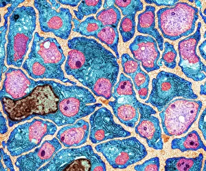
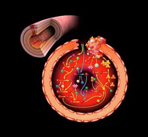
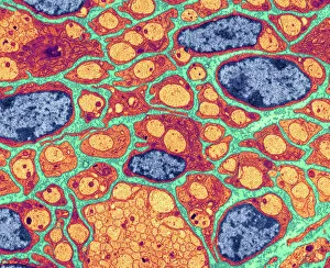
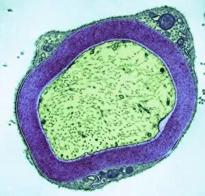
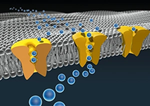
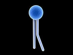
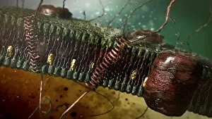
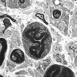
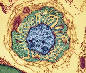
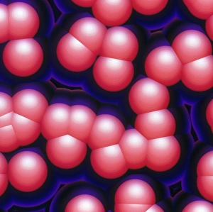
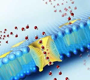
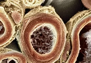
Phospholipids: The Dynamic Builders of Life's Blueprint From the intricate cell membrane to the myelination of nerve fibers, phospholipids play a vital role in various biological processes. In artwork C013/7467, we witness their remarkable presence as they form the foundation of the cell membrane, providing structure and stability. Moving on to TEM images showcasing myelination of nerve fibers, we observe how phospholipids contribute to this crucial process. These lipid molecules wrap around nerve fibers like a protective sheath (TEM image), ensuring efficient transmission of electrical signals throughout our body. But that's not all. Phospholipids also participate in blood coagulation cascade (artwork C016/9873). They interact with proteins involved in clotting, facilitating this essential mechanism that prevents excessive bleeding. Returning to TEM images once again, we explore how phospholipids enable ion channels within cell membranes (artwork C016/7689). These specialized pathways allow ions to flow across cellular boundaries, regulating important physiological functions such as muscle contraction and nerve signaling. In conceptual imagery depicting lipids (F006/9780), we gain insight into their diverse roles beyond structural support. Phospholipids serve as energy stores and act as messengers within cells - orchestrating complex biochemical reactions necessary for life itself. As we zoom back into specific artworks highlighting phospholipid arrangements within membranes (C018/7905), it becomes evident that these molecules are intricately organized. Their unique properties ensure selective permeability while maintaining cellular integrity - an extraordinary feat achieved by nature's design. Finally, let us marvel at individual phospholipid molecules depicted in stunning artwork. With their hydrophilic heads and hydrophobic tails perfectly balanced, they create a harmonious arrangement critical for proper functioning at a microscopic level. Last but not least, TEM images reveal the importance of phospholipids in nerve fiber nodes.