
Ovaries Collection
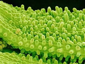
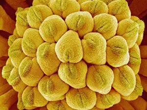
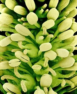
"Exploring the Intricacies of Ovaries: Unveiling Nature's Masterpieces" Delving into the realm of reproductive wonders, we encounter the captivating Euphorbia flower


All Professionally Made to Order for Quick Shipping
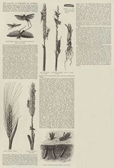
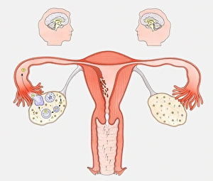
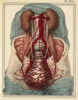
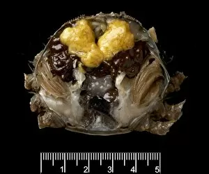
"Exploring the Intricacies of Ovaries: Unveiling Nature's Masterpieces" Delving into the realm of reproductive wonders, we encounter the captivating Euphorbia flower. Its intricate reproductive parts, resembling delicate brushstrokes on a canvas, showcase nature's artistry. Through scanning electron microscopy (SEM), we witness a mesmerizing front view of the female anatomy, unveiling not only its physicality but also highlighting its vital role in the endocrine system. Journeying back in time to 1830, an exquisite painted cardboard depiction of female genitals emerges. This artwork from centuries past reminds us of our ancestors' fascination with understanding and appreciating the intricacies of human reproduction. The woodcut illustration by Johannes de Ketham in 1491 takes us further into history as it portrays "The Anatomy of the Pregnant Woman. " This ancient representation captures both scientific curiosity and artistic expression, offering insights into how pregnancy was perceived during that era. Venturing beyond human biology, an engraving depicting "The Plague of Crickets in Algeria" serves as a reminder that nature's cycles extend far beyond our own existence. It prompts contemplation about life's interconnectedness and how various organisms navigate their own reproductive journeys. Returning to human reproduction systems through another woodcut from 1554, we observe a pregnant woman's intricate internal structure. The detailed portrayal invites reflection on the remarkable processes occurring within a woman's body during this transformative period. A comprehensive diagram showcases not only the interaction between female sexual organs and the brain but also presents contrasting perspectives – one illustrating normal reproductive cycles while juxtaposed with another demonstrating contraceptive pill effects. Such visual aids deepen our understanding of fertility control methods throughout history and highlight advancements made for women worldwide. Transitioning to menopause, conceptual artwork evokes emotions surrounding this significant phase in every woman's life journey. Symbolically capturing both endings and new beginnings, it celebrates resilience while acknowledging the complexities of this natural transition.