Fraud Blocker Skip to main content

Nerve Collection (page 6)

"Nerve: Unveiling the Intricacies of the Human Brain and its Pathways" Delving into the depths of our complex neural network

Background imageNerve Collection: Central nervous system with human brain

Central nervous system with human brain

Background imageNerve Collection: Conceptual image of a neuron ligntning signal passing

Conceptual image of a neuron ligntning signal passing

Background imageNerve Collection: Medical illustration of the human nervous system and brain

Medical illustration of the human nervous system and brain, front view

Background imageNerve Collection: Neurofibromatosis, a genetic disorder of the nervous system

Neurofibromatosis, a genetic disorder of the nervous system
A genetic disorder of the nervous system, neurofibromatosis causes tumors to form on nerves throughout the body, including a type of tumor called an optic nerve glioma that can result in childhood

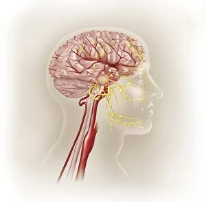
Background imageNerve Collection: Detail of ateries of the human head and the trigeminal nerve

Detail of ateries of the human head and the trigeminal nerve

Background imageNerve Collection: Medical illustration of human arm muscles, veins and nerves

Medical illustration of human arm muscles, veins and nerves

Background imageNerve Collection: Microscopic view of inner nervous system

Microscopic view of inner nervous system

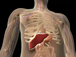
Background imageNerve Collection: Transparent view of human torso showing liver

Transparent view of human torso showing liver

Background imageNerve Collection: Illustration of action potential of a nerve cell

Illustration of action potential of a nerve cell

Background imageNerve Collection: Conceptual image of a neuron

Conceptual image of a neuron

Background imageNerve Collection: Medical illustration of human brain and brain stem

Medical illustration of human brain and brain stem, perspective view

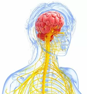
Background imageNerve Collection: Anatomy of female body with nervous system

Anatomy of female body with nervous system

Background imageNerve Collection: Cross section of human ear

Cross section of human ear

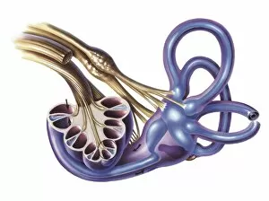
Background imageNerve Collection: Cochlea detail with vestibulocochlear nerve

Cochlea detail with vestibulocochlear nerve

Background imageNerve Collection: External auditory canal of human ear

External auditory canal of human ear

Background imageNerve Collection: Right side view of the brain and cerebellum. Woodcut from the fourth book of Andreas Vesalius De

Right side view of the brain and cerebellum. Woodcut from the fourth book of Andreas Vesalius De Humani Corporis
BRAIN & CEREBELLUM, 1543. Right side view of the brain and cerebellum. Woodcut from the fourth book of Andreas Vesalius De Humani Corporis Fabrica, published in 1543 at Basel, Switzerland

Background imageNerve Collection: Delineation of the spinal nerves. Woodcut from the fourth book of Andreas Vesalius De Humani

Delineation of the spinal nerves. Woodcut from the fourth book of Andreas Vesalius De Humani Corporis Fabrica
VESALIUS: NERVES, 1543. Delineation of the spinal nerves. Woodcut from the fourth book of Andreas Vesalius De Humani Corporis Fabrica, published in 1543 at Basel, Switzerland

Background imageNerve Collection: The base of the entire brain and cerebellum freed from the surrounding membranes so that

The base of the entire brain and cerebellum freed from the surrounding membranes so that the origin of the cerebral
VESALIUS: BRAIN. The base of the entire brain and cerebellum freed from the surrounding membranes so that the origin of the cerebral nerves are exposed to view

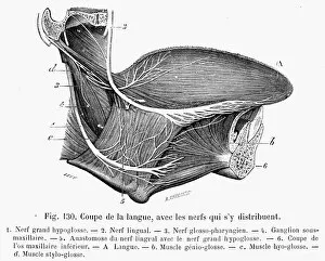
Background imageNerve Collection: Nerves distributed within the tongue. Line engraving, French, 19th century

Nerves distributed within the tongue. Line engraving, French, 19th century
HUMAN TONGUE AND NERVES. Nerves distributed within the tongue. Line engraving, French, 19th century

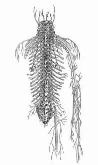
Background imageNerve Collection: Posterior aspect of the spinal nerves. Woodcut from the fourth book of Andreas Vesaliuss De Humani

Posterior aspect of the spinal nerves. Woodcut from the fourth book of Andreas Vesaliuss De Humani Corporis Fabrica
ANATOMY: SPINAL NERVES. Posterior aspect of the spinal nerves. Woodcut from the fourth book of Andreas Vesaliuss De Humani Corporis Fabrica, Basel, 1543
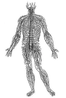
Background imageNerve Collection: Woodcut from the fourth book of Andreas Vesalius De Humani Corporis Fabrica

Woodcut from the fourth book of Andreas Vesalius De Humani Corporis Fabrica, published in 1543 at Basel, Switzerland
VESALIUS: NERVES, 1543. Woodcut from the fourth book of Andreas Vesalius De Humani Corporis Fabrica, published in 1543 at Basel, Switzerland

Background imageNerve Collection: Albrecht von Haller (1708-1777), Swiss physician and scientist who was the founder of neurology

Albrecht von Haller (1708-1777), Swiss physician and scientist who was the founder of neurology, c1770. Experimental physiology. Engraving c1790

Background imageNerve Collection: Diagram illustrating Descartes idea of vision, showing the function of the eye

Diagram illustrating Descartes idea of vision, showing the function of the eye, optic nerve and brain. From Rene Descartes Opera Philiosophica, Frankfurt-am-Main, 1692. Woodcut

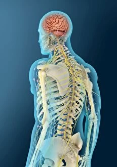
Background imageNerve Collection: Illustration showing human nervous system

Illustration showing human nervous system
Medicine: nervous system

Background imageNerve Collection: Illustration of human nervous system

Illustration of human nervous system
Medicine: nervous system. Drawing

Background imageNerve Collection: Illustration of human eye with muscles, blood vessels and nerves

Illustration of human eye with muscles, blood vessels and nerves
Medicine: Human anatomy, eye. Muscles, blood vessels and nerves

Background imageNerve Collection: Illustration of periodontium

Illustration of periodontium
Medicine: Human anatomy, periodontium

Background imageNerve Collection: Illustration of extrapyramidal system

Illustration of extrapyramidal system
Medicine: Human anatomy, extrapyramidal system. Drawing

Background imageNerve Collection: Illustration of sensibility to physical pain

Illustration of sensibility to physical pain
Medicine: Human anatomy, sensibility to physical pain

Background imageNerve Collection: Illustration of abducens nerve, or sixth cranial nerve

Illustration of abducens nerve, or sixth cranial nerve
Medicine: Human anatomy, abducens nerve, or sixth cranial nerve. Drawing

Background imageNerve Collection: Illustration of human spinal accessory nerve

Illustration of human spinal accessory nerve
Medicine: Human anatomy, spinal accessory nerve. Drawing

Background imageNerve Collection: Illustration of spinal cord

Illustration of spinal cord
Medicine: Human anatomy, spinal cord. Drawing

Background imageNerve Collection: Overhead view of model of upper torso of Giant Tiger Centipede

Overhead view of model of upper torso of Giant Tiger Centipede, pair of legs attached to each segment, venom claws behind antennae, cross-section showing venom glands with duct, nerve and muscle

Background imageNerve Collection: Electric ray brain, illustration C018 / 0913

Electric ray brain, illustration C018 / 0913
Electric ray brain, illustration. Electric rays are able to produce an electric discharge, to stun prey or for defence, from two electric organs situated either side of their heads

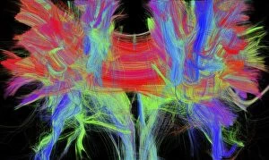
Background imageNerve Collection: Corpus callosum, DTI MRI scan C017 / 7044

Corpus callosum, DTI MRI scan C017 / 7044
Corpus callosum. Coronal 3D diffusion tensor imaging (DTI) magnetic resonance imaging (MRI) scan of nerve pathways (coloured) in and around the brains corpus callosum

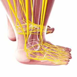
Background imageNerve Collection: Human foot nervous system, artwork F007 / 1148

Human foot nervous system, artwork F007 / 1148
Human foot nervous system, computer artwork

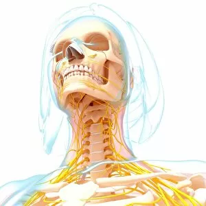
Background imageNerve Collection: Female nervous system, artwork F007 / 4284

Female nervous system, artwork F007 / 4284
Female nervous system, computer artwork

Background imageNerve Collection: Female nervous system, artwork F007 / 3461

Female nervous system, artwork F007 / 3461
Female nervous system, computer artwork

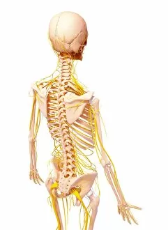
Background imageNerve Collection: Human nervous system, artwork F007 / 3986

Human nervous system, artwork F007 / 3986
Human nervous system, computer artwork

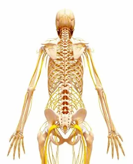
Background imageNerve Collection: Human nervous system, artwork F007 / 3394

Human nervous system, artwork F007 / 3394
Human nervous system, computer artwork

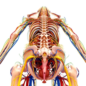
Background imageNerve Collection: Female anatomy, artwork F007 / 3969

Female anatomy, artwork F007 / 3969
Female anatomy, computer artwork

Background imageNerve Collection: Female anatomy, artwork F007 / 3665

Female anatomy, artwork F007 / 3665
Female anatomy, computer artwork

Background imageNerve Collection: Human nervous system, artwork F007 / 4283

Human nervous system, artwork F007 / 4283
Human nervous system, computer artwork

Background imageNerve Collection: Female nervous system, artwork F007 / 4298

Female nervous system, artwork F007 / 4298
Female nervous system, computer artwork

Background imageNerve Collection: Human nervous system, artwork F007 / 4193

Human nervous system, artwork F007 / 4193
Human nervous system, computer artwork

Background imageNerve Collection: Female anatomy, artwork F007 / 4084

Female anatomy, artwork F007 / 4084
Female anatomy, computer artwork

Background imageNerve Collection: Female anatomy, artwork F007 / 3976

Female anatomy, artwork F007 / 3976
Female anatomy, computer artwork


All Professionally Made to Order for Quick Shipping

"Nerve: Unveiling the Intricacies of the Human Brain and its Pathways" Delving into the depths of our complex neural network, this captivating caption takes us on a journey through the fascinating world of nerves. From brain fibres to DTI MRI scans (C017 / 7099) that reveal intricate connections, we explore the anatomy of the human brain from an inferior view. As we unravel the mysteries within, nerve and glial cells come to light under a microscope's gaze in a mesmerizing light micrograph. The Spanish histologist Santiago Ramon y Cajal's meticulous drawing from 1894 showcases various cell types in the mammalian cerebellum, reminding us of how far our understanding has evolved. The cross-section diagram guides us towards another vital aspect - our auditory system. Exploring a detailed illustration of both ear and brain reveals how nerves play an integral role in transmitting sound signals for perception. Zooming further into microscopic wonders, we encounter individual nerve cells with their intricate structures and functions. These remarkable entities are responsible for relaying information throughout our body, allowing us to experience sensations and perform actions effortlessly. With every piece falling into place like a puzzle, an illustration depicting both spinal cord and brain highlights their interconnectedness. This visual representation emphasizes how nerves form an essential bridge between these two crucial components of our central nervous system. Amidst this scientific exploration lies unexpected nostalgia as we stumble upon an Ovaltine Advert – perhaps reminding us that even everyday experiences rely on well-functioning nerves. Finally, hope arises as we witness regenerating nerve cells through transmission electron microscopy (TEM). This image encapsulates nature's ability to heal itself—a testament to resilience within our intricate neural framework. "Nerve" unravels layers upon layers of complexity surrounding one fundamental element that defines human existence—the delicate yet powerful web connecting every fiber within ourselves -our nerves.