
Nerve Collection (page 4)
"Nerve: Unveiling the Intricacies of the Human Brain and its Pathways" Delving into the depths of our complex neural network


All Professionally Made to Order for Quick Shipping
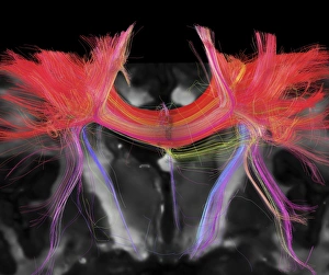
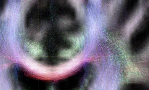
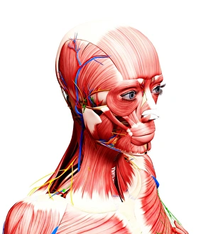
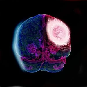
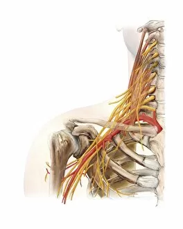
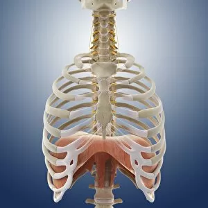
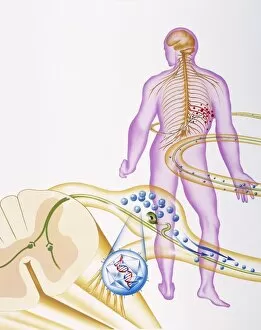
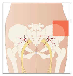
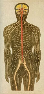
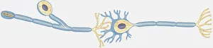
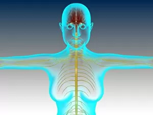
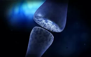
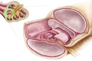
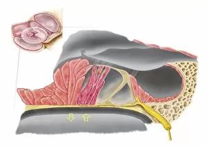
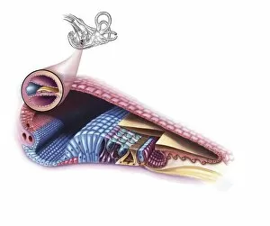
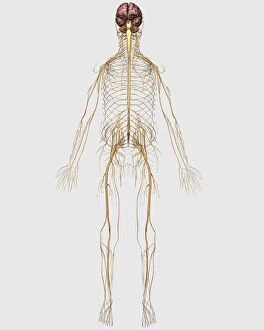
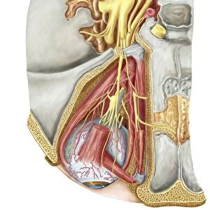
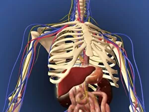
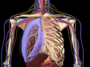
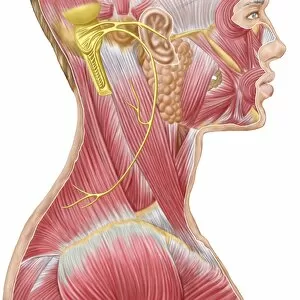
"Nerve: Unveiling the Intricacies of the Human Brain and its Pathways" Delving into the depths of our complex neural network, this captivating caption takes us on a journey through the fascinating world of nerves. From brain fibres to DTI MRI scans (C017 / 7099) that reveal intricate connections, we explore the anatomy of the human brain from an inferior view. As we unravel the mysteries within, nerve and glial cells come to light under a microscope's gaze in a mesmerizing light micrograph. The Spanish histologist Santiago Ramon y Cajal's meticulous drawing from 1894 showcases various cell types in the mammalian cerebellum, reminding us of how far our understanding has evolved. The cross-section diagram guides us towards another vital aspect - our auditory system. Exploring a detailed illustration of both ear and brain reveals how nerves play an integral role in transmitting sound signals for perception. Zooming further into microscopic wonders, we encounter individual nerve cells with their intricate structures and functions. These remarkable entities are responsible for relaying information throughout our body, allowing us to experience sensations and perform actions effortlessly. With every piece falling into place like a puzzle, an illustration depicting both spinal cord and brain highlights their interconnectedness. This visual representation emphasizes how nerves form an essential bridge between these two crucial components of our central nervous system. Amidst this scientific exploration lies unexpected nostalgia as we stumble upon an Ovaltine Advert – perhaps reminding us that even everyday experiences rely on well-functioning nerves. Finally, hope arises as we witness regenerating nerve cells through transmission electron microscopy (TEM). This image encapsulates nature's ability to heal itself—a testament to resilience within our intricate neural framework. "Nerve" unravels layers upon layers of complexity surrounding one fundamental element that defines human existence—the delicate yet powerful web connecting every fiber within ourselves -our nerves.