
Myology Collection (page 4)
Myology: Exploring the Intricacies of Human Muscles From the delicate facial muscles that shape our expressions to the powerful ligaments that support our knees


All Professionally Made to Order for Quick Shipping
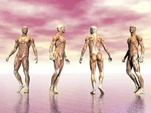
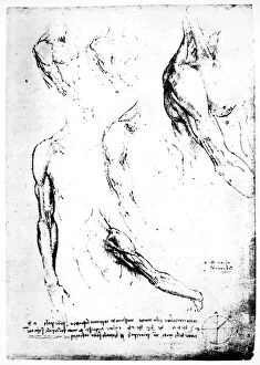
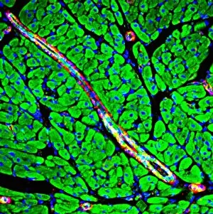
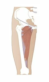
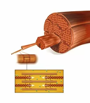
Myology: Exploring the Intricacies of Human Muscles From the delicate facial muscles that shape our expressions to the powerful ligaments that support our knees, myology delves into the fascinating world of human musculature. Intricate and interconnected, the facial muscles of the human face are a marvel to behold. With labels guiding us through their complexity, we gain insight into how these muscles work together to create smiles, frowns, and everything in between. A damaged knee ligament serves as a reminder of both the vulnerability and resilience of our bodies. Through artwork capturing this injury, we witness firsthand the importance of proper care and rehabilitation for such crucial structures. Julien Bougle's masterpiece presents us with an awe-inspiring visualization - colored plates superimposed on the human body. This artistic representation allows us to appreciate not only individual muscle groups but also their relationship with other internal organs. The eye muscle is a remarkable example of precision engineering within our bodies. TEM C014 / 1468 showcases its intricate structure, reminding us just how intricately designed even seemingly small muscles can be. Artwork depicting human muscle structure takes us on a journey beneath our skin's surface. We marvel at each fiber and tendon working harmoniously to enable movement and strength throughout our entire being. Shoulder muscles play a vital role in maintaining upper body mobility and stability. Artistic renderings allow us to explore their form and function while appreciating their contribution to everyday activities like lifting or reaching overhead. Anatomy lessons extend beyond superficial appearances; they delve deep into every part of our bodies - even those hidden from plain sight. The gluteal muscles in our buttocks reveal themselves under scrutiny as essential contributors to posture, balance, and locomotion. As we venture further inside ourselves, exploring musculature alongside internal organs becomes an enlightening experience indeed. Understanding how these systems coexist enables appreciation for both their complexity and the remarkable harmony they achieve.