
Musculoskeletal Disorders Collection
Musculoskeletal disorders can greatly impact our daily lives, causing pain and limiting our mobility


All Professionally Made to Order for Quick Shipping
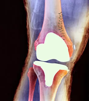
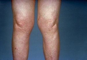
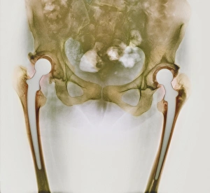
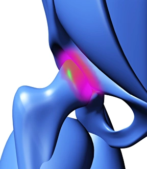
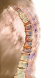
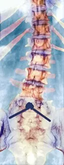
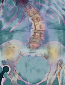
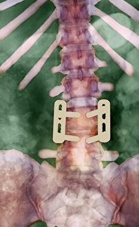
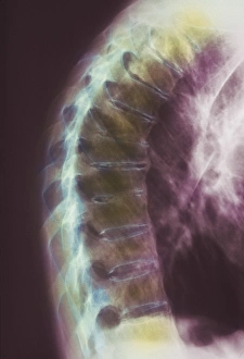
Musculoskeletal disorders can greatly impact our daily lives, causing pain and limiting our mobility. From knee joint prostheses to X-rays revealing pinned vertebrae, these images shed light on the challenges faced by individuals with musculoskeletal conditions. In one snapshot, we see a view of knees affected by osteoarthritis, showcasing the degenerative nature of this disease. The X-ray reveals the toll it takes on the joints, highlighting the need for effective treatment options. Another image captures an arthritic hand through an X-ray lens. The intricate bones show signs of wear and tear caused by inflammation and joint damage. These visuals remind us of the importance of early intervention in managing arthritis. Osteoporosis is another common musculoskeletal disorder that weakens bones over time. Through X-rays labeled C017/8250, 8248, 8245, and 8244 respectively, we witness how this condition affects bone density and increases fracture risk. It serves as a reminder to prioritize bone health throughout our lives. Amidst these medical depictions lies a moment of relief - a woman receiving a neck and shoulder massage. This therapeutic touch offers respite from muscle tension often associated with musculoskeletal disorders. Lastly, another glimpse into knee joint prostheses via X-ray showcases advancements in medical technology aimed at restoring mobility for those suffering from severe joint damage. These snapshots provide insight into various aspects - their effects on different body parts like knees or hands; their presence in conditions such as osteoarthritis or osteoporosis; and potential interventions like massages or prosthetic replacements. By understanding these complexities better, we can work towards improving prevention strategies and enhancing treatments for individuals living with musculoskeletal disorders.