
Mitochondrial Collection
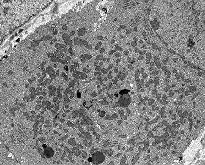
Mitochondrial, the powerhouse of our cells, plays a crucial role in energy production and various cellular processes


All Professionally Made to Order for Quick Shipping
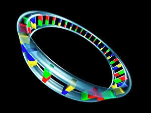
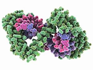
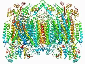
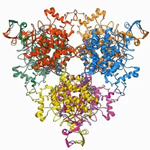
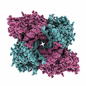
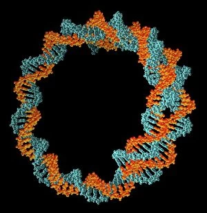
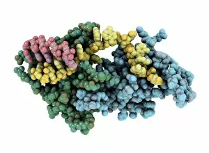
Mitochondrial, the powerhouse of our cells, plays a crucial role in energy production and various cellular processes. At its core lies mitochondrial DNA (mtDNA), which encodes essential genes for oxidative phosphorylation. The ATPase molecule, an integral part of this process, generates adenosine triphosphate (ATP) - the currency of cellular energy. TFAM transcription factor bound to DNA C015 / 7059 regulates mtDNA replication and transcription, ensuring proper maintenance and function. Meanwhile, the Citrate acid cycle enzyme F006 / 9305 initiates a series of reactions that extract high-energy electrons from nutrients. SIRT3 molecule, artwork C017 / 3657 acts as a guardian health by regulating protein acetylation levels and maintaining optimal mitochondrial function. The ATP-binding cassette transporter F006 / 9743 facilitates the transport of molecules across mitochondrial membranes while Beta-lactamase-like protein 2 molecule F006 / 9741 contributes to antibiotic resistance within mitochondria. Single stranded DNA-binding protein F006 / 9733 protects mtDNA during replication and repair processes. Additionally, Citrate synthase molecule F006 /9573 catalyzes a key step in the citric acid cycle. Cytochrome c oxidase and antibody F006/9474 are involved in electron transfer within mitochondria's respiratory chain. This vital process ultimately leads to ATP synthesis. P32 mitochondrial matrix protein F006/9454 participates in RNA processing within mitochondria while Cytochrome c oxidase molecule F006/9447 is an essential component responsible for transferring electrons to oxygen during oxidative phosphorylation. These intricate components collectively contribute to maintaining efficient energy production and overall cellular homeostasis within our mighty mitochondria.