
Male Reproductive System Collection
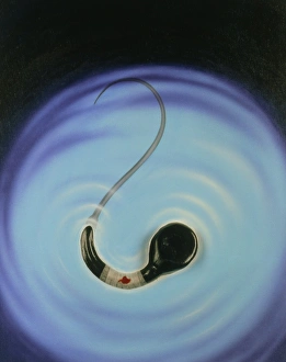
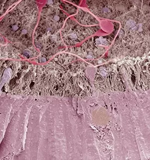
"Exploring the Intricacies of the Male Reproductive System: Unveiling its Anatomy and Functions" Deformed sperm cell under SEM


All Professionally Made to Order for Quick Shipping
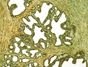
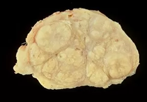
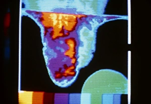
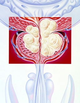
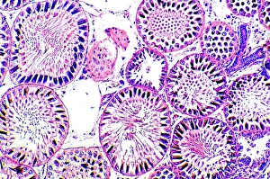
"Exploring the Intricacies of the Male Reproductive System: Unveiling its Anatomy and Functions" Deformed sperm cell under SEM: A closer look at the intricate world of male reproductive cells, revealing fascinating abnormalities. Anatomy of male urinary bladder, anterior view: Delving into the structure and positioning of the vital organ responsible for storing urine in males. Sagittal view of male urinary bladder: Peering inside to understand how this remarkable organ functions within the male reproductive system. Conceptual image of human male reproductive organs: Unlocking a visual representation that showcases the complexity and interconnectedness of these essential components. 3D rendering of digestive system and male reproductive system: Merging two crucial bodily systems to highlight their interplay in maintaining overall health and functionality. Biomedical illustration of a vasectomy and tubectomy: Shedding light on surgical procedures aimed at permanent contraception, empowering individuals with choices regarding family planning. 3D rendering of human skeleton with internal organs: Offering an immersive perspective by showcasing how bones support and protect vital structures like those found in the male reproductive system. Anatomy of male and female urinary bladder: Comparing both genders' bladder anatomy to appreciate similarities as well as differences between their respective systems. Anterior view and sagittal view of male urinary bladder: Examining multiple angles to gain comprehensive insights into this key component's location within males' lower abdominal region. Conceptual image (s)of human male reproductive organs (repeated).