
Lyme Disease Collection
"Lyme Disease: Unveiling the Silent Intruder" In 1906, a groundbreaking discovery was made when a lithograph revealed the presence of Spirochaetes Borrelia Recurrentis


All Professionally Made to Order for Quick Shipping
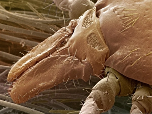
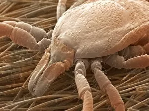
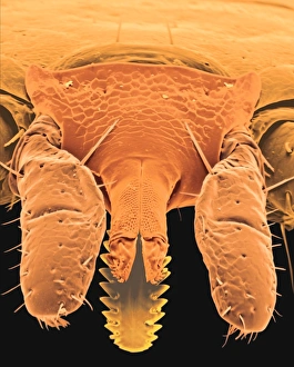
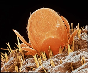
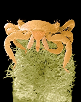
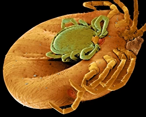
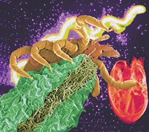
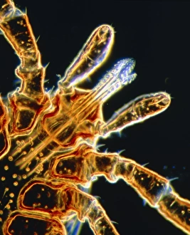
"Lyme Disease: Unveiling the Silent Intruder" In 1906, a groundbreaking discovery was made when a lithograph revealed the presence of Spirochaetes Borrelia Recurrentis, the bacteria responsible for Lyme disease. This microscopic view showcased the intricate spirillum structure of these elusive culprits lurking within blood samples. Fast forward to today, and we now have advanced technology allowing us to witness Borrelia burgdorferi bacteria in all their glory. These captivating images captured through SEM reveal clusters of these tiny organisms, resembling an army ready to invade. But how does this stealthy invasion occur? Enter the deer tick and sheep tick - carriers of this debilitating disease. SEM images showcase their minuscule bodies with astonishing detail, highlighting their role as vectors for transmitting Lyme disease. These ticks are not limited by habitat; they can be found on plant leaves or even embedded in human skin. Colored SEM images vividly portray these encounters - from ticks feeding on unsuspecting legs to Ixodes sp. Firmly latching onto plants or humans alike. Lyme disease is more than just a mere nuisance; it poses significant health risks if left untreated. Awareness is key in preventing its spread and ensuring early diagnosis for affected individuals. So let us join forces against this silent intruder. By understanding its transmission through ticks like never before thanks to SEM imaging, we can take proactive measures such as wearing protective clothing and conducting regular tick checks after outdoor activities. Remember, knowledge empowers us in our fight against Lyme disease – together we can protect ourselves and loved ones from its grasp.