Fraud Blocker Skip to main content

Liver Cell Collection

The liver cell, also known as a hepatocyte, plays a crucial role in maintaining our overall health

Background imageLiver Cell Collection: Hepatitis C viruses, TEM

Hepatitis C viruses, TEM
Hepatitis C viruses. Coloured transmission electron micrograph (TEM) of hepatitis C virus particles (green) infecting cultured liver cells (purple)

Background imageLiver Cell Collection: Golgi apparatus, SEM

Golgi apparatus, SEM
Golgi apparatus, coloured scanning electron micrograph (SEM). Section through a liver cell showing its Golgi apparatus (grey), a membrane-bound organelle that modifies and packages proteins

Background imageLiver Cell Collection: False-colour TEM of a human hepatocyte

False-colour TEM of a human hepatocyte
False colour transmission electron micrograph (TEM) of a section through a human hepatocyte, a liver cell. Hepatocytes function in the storage of glycogen (metabolized from excess glucose)

Background imageLiver Cell Collection: Coloured SEM of liver cells and bile canaliculi

Coloured SEM of liver cells and bile canaliculi

Background imageLiver Cell Collection: False-colour TEM of a section through a liver cell

False-colour TEM of a section through a liver cell
False colour transmission electron micrograph (TEM) of a section through a human hepatocyte, a liver cell. Hepatocytes function in the storage of glycogen (metabolized from excess glucose)

Background imageLiver Cell Collection: False-colour SEM of vessels in liver lobule

False-colour SEM of vessels in liver lobule
Liver tissue. False-colour scanning electron micrograph (SEM) of blood vessels around a lobule, the functional unit of the liver

Background imageLiver Cell Collection: Liver cells, light micrograph

Liver cells, light micrograph
Liver cells. Light micrograph of a section through a human liver. The liver cells are pink with purple nuclei. A duct canal and its surrounding tissue can be seen at lower left

Background imageLiver Cell Collection: Illustration of structure of human hepatocyte

Illustration of structure of human hepatocyte
Artwork illustrating the compact internal structure of a human hepatocyte, a liver cell. The cytoplasm of a hepatocyte is crowded with organelles; the density relects its numerous metabolic

Background imageLiver Cell Collection: Coloured SEM of a bile canaliculus in the liver

Coloured SEM of a bile canaliculus in the liver
Bile canaliculus. Coloured scanning electron micrograph of a human liver cell (red-brown), also known as hepatocyte, and a bile canaliculus (green)

Background imageLiver Cell Collection: Liver cells, TEM

Liver cells, TEM
Liver cells. Coloured transmission electron micrograph (TEM) of cells in a liver, showing their tessellating boundaries (red) and nuclei (beige). Magnification: x9000 when printed 10 centimetres wide

Background imageLiver Cell Collection: Colour TEM of primary lysosome in liver cell

Colour TEM of primary lysosome in liver cell
Primary lysosome. Coloured Transmission Electron Micrograph (TEM) of a primary lysosome seen in a liver cell. The lysosome (red) is a membrane bound structure that contains digestive enzymes

Background imageLiver Cell Collection: Coloured SEM of mitochondria & ER in a liver cell

Coloured SEM of mitochondria & ER in a liver cell
Mitochondria. Coloured Scanning Electron Micro- graph (SEM) of mitochondria and rough endoplasmic reticulum in a liver cell


All Professionally Made to Order for Quick Shipping

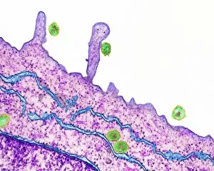
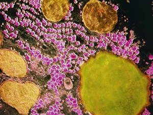
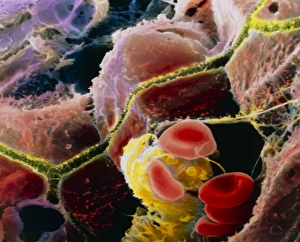
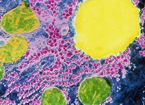
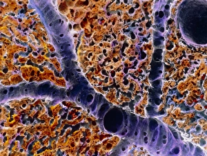
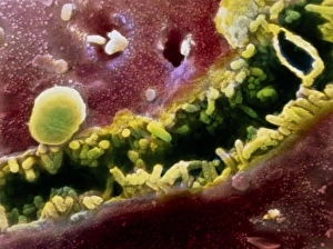
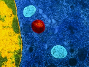
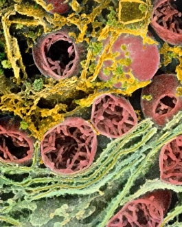
The liver cell, also known as a hepatocyte, plays a crucial role in maintaining our overall health. These remarkable cells are involved in various processes that keep our body functioning optimally. One significant challenge faced by liver cells is the invasion of Hepatitis C viruses. Through the use of Transmission Electron Microscopy (TEM), scientists have been able to observe these tiny viral particles interacting with hepatocytes at an incredibly detailed level. Another essential organelle found within liver cells is the Golgi apparatus. Scanning Electron Microscopy (SEM) has allowed researchers to visualize this complex structure and understand its role in processing and packaging proteins for secretion. In a False-colour TEM image of a human hepatocyte, we can appreciate the intricate network of cellular components that make up these vital cells. The image showcases their unique morphology and highlights specific organelles such as mitochondria and endoplasmic reticulum. Coloured SEM images provide us with an even more vivid representation of liver cells and their surrounding environment. In one striking image, we can see liver cells along with bile canaliculi - small ducts responsible for transporting bile produced by hepatocytes. A section through a liver cell captured using False-colour TEM reveals further details about its internal structure. This high-resolution image allows us to study different compartments within the cell, including nuclei and cytoplasmic components like ribosomes. Within each lobule of the liver lies an intricate network of vessels that ensure proper blood supply for metabolic functions. A captivating False-colour SEM image beautifully illustrates this vascular system within a liver lobule. When observed under light microscopy, individual hepatocytes appear as distinct polygonal shapes forming tissue patterns characteristic of healthy livers. This light micrograph provides valuable insights into their arrangement within hepatic tissue sections. An illustration depicting the structure of a human hepatocyte offers an informative overview highlighting key features such as nuclei, mitochondria, Golgi apparatus, and rough endoplasmic reticulum.