Fraud Blocker Skip to main content

Larynx Collection (page 5)

The larynx, also known as the voice box, is a vital part of the human respiratory system and can be seen in an illustration depicting the entire respiratory system

Background imageLarynx Collection: Female lungs, artwork

Female lungs, artwork
Female lungs, computer artwork

Background imageLarynx Collection: Male anatomy, artwork F006 / 8353

Male anatomy, artwork F006 / 8353
Male anatomy, computer artwork

Background imageLarynx Collection: Healthy respiratory system, artwork F006 / 7841

Healthy respiratory system, artwork F006 / 7841
Healthy respiratory system, computer artwork

Background imageLarynx Collection: Larynx cancer, artwork

Larynx cancer, artwork
Larynx cancer, computer artwork

Background imageLarynx Collection: Female anatomy, artwork F006 / 7796

Female anatomy, artwork F006 / 7796
Female anatomy, computer artwork

Background imageLarynx Collection: Female anatomy, artwork F006 / 7773

Female anatomy, artwork F006 / 7773
Female anatomy, computer artwork

Background imageLarynx Collection: Female anatomy, artwork F006 / 7766

Female anatomy, artwork F006 / 7766
Female anatomy, computer artwork

Background imageLarynx Collection: Female anatomy, artwork F006 / 7761

Female anatomy, artwork F006 / 7761
Female anatomy, computer artwork

Background imageLarynx Collection: Female anatomy, artwork F006 / 7757

Female anatomy, artwork F006 / 7757
Female anatomy, computer artwork

Background imageLarynx Collection: Lung cancer, artwork

Lung cancer, artwork
Lung cancer, computer artwork

Background imageLarynx Collection: Voice box anatomy, artwork C016 / 7692

Voice box anatomy, artwork C016 / 7692
Voice box anatomy. Computer artwork showing the location (right) and structure (left) of a human larynx (voice box). The larynx is involved in breathing, sound production (phonation)

Background imageLarynx Collection: Oedema of larynx, artwork C016 / 6529

Oedema of larynx, artwork C016 / 6529
Oedema of larynx. Artwork of a transverse (axial) section through a larynx (seen from above) with and without swelling. The bottom image shows a healthy larynx

Background imageLarynx Collection: Oedema of larynx, artwork C016 / 6528

Oedema of larynx, artwork C016 / 6528
Oedema of larynx. Artwork of a vertical (coronal) section through a larynx, showing oedema (swelling caused by fluid accumulation)

Background imageLarynx Collection: Oedema of larynx, artwork C016 / 6530

Oedema of larynx, artwork C016 / 6530
Oedema of larynx. Artwork of sections through a larynx with and without swelling. The bottom image shows a healthy larynx in transverse (axial) section from above

Background imageLarynx Collection: Human throat anatomy, artwork

Human throat anatomy, artwork
Human throat anatomy. Computer artwork of a human throat, showing the oesophagus (gullet, red, vertical left) and trachea (windpipe, red vertical right)

Background imageLarynx Collection: Healthy larynx and thyroid, artwork C014 / 0402

Healthy larynx and thyroid, artwork C014 / 0402
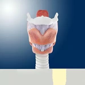
Healthy larynx and thyroid, computer artwork. At centre is the larynx (purple/grey), also known as the voice box, surrounded by the thyroid cartilage (pink).Above the larynx is the hyoid bone (white)

Background imageLarynx Collection: Healthy larynx and thyroid, artwork

Healthy larynx and thyroid, artwork
Healthy larynx and thyroid, computer artwork. At centre is the larynx (purple/grey), also known as the voice box, surrounded by the thyroid cartilage (pink)

Background imageLarynx Collection: Healthy larynx and thyroid, artwork C014 / 0404

Healthy larynx and thyroid, artwork C014 / 0404
Healthy larynx and thyroid, computer artwork. The larynx, also known as the voice box, is purple/grey. It is surrounded by the thyroid cartilage (pink)

Background imageLarynx Collection: Healthy larynx and thyroid, artwork C014 / 0400

Healthy larynx and thyroid, artwork C014 / 0400
Healthy larynx and thyroid, computer artwork. At centre is the larynx (purple/grey), also known as the voice box, surrounded by the thyroid cartilage (pink)

Background imageLarynx Collection: Healthy larynx, artwork C014 / 0403

Healthy larynx, artwork C014 / 0403
Healthy larynx, computer artwork. At centre is the larynx (purple/grey), also known as the voice box, surrounded by the thyroid cartilage (pink)

Background imageLarynx Collection: Human throat, anatomical model

Human throat, anatomical model
Human throat. Anatomical model of the human throat, showing the teeth of the lower jaw, voicebox (larynx, beige, centre), and windpipe (trachea, pink tube)

Background imageLarynx Collection: Lungs and diaphragm, 1825 artwork

Lungs and diaphragm, 1825 artwork
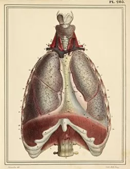
Lungs and diaphragm. Anterior view of the chest, dissected vertically to reveal the lobes of the lungs, the diaphragm (brown, across bottom)

Background imageLarynx Collection: Diseased organs, artwork

Diseased organs, artwork
Diseased organs. Computer artwork showing diseased major organs of the human torso. From top to bottom: larynx (voicebox), trachea (windpipe), lungs (centre left and right), heart (centre), liver

Background imageLarynx Collection: Heart and lungs, artwork

Heart and lungs, artwork
Heart and lungs. Computer artwork of the heart and bronchial trees of the lungs seen from below. At upper right is the trachea (windpipe), which splits into two bronchi, one for each lung

Background imageLarynx Collection: Respiratory system, artwork C015 / 6453

Respiratory system, artwork C015 / 6453
Respiratory system. Computer artwork showing the organs of the human respiratory system. At top is the larynx (voicebox), which leads onto the trachea (windpipe, white tube vertical)

Background imageLarynx Collection: Thyroid anatomy, artwork C013 / 4674

Thyroid anatomy, artwork C013 / 4674
Thyroid anatomy. Computer artwork of the thyroid glands (pink) of the neck, shown in relation to the bodys bones and throat structures (white)

Background imageLarynx Collection: WW2 - Blitz, London - Progress report sent to Local Control

WW2 - Blitz, London - Progress report sent to Local Control
Senior Company Officer Jimmy Syrett, up with the branchmen, sends a progress report to Local Control. He is using one of the early walkie talkies with a throat, or larynx, microphone

Background imageLarynx Collection: Anatomy / Larynx / Diaphragm

Anatomy / Larynx / Diaphragm
Muscles of the diaphragm and the larynx according to the anatomists Haller and Eustachi Date: Circa 1760

Background imageLarynx Collection: Artwork of the nose, mouth and throat in profile

Artwork of the nose, mouth and throat in profile
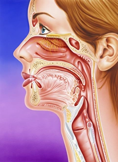
Nose, mouth and throat. Artwork of a human face in profile showing detail of the mouth, nose and throat. Leading back from the nose (upper left) is the nasal cavity

Background imageLarynx Collection: Nose, mouth and throat

Nose, mouth and throat. Cutaway artwork of the anatomy of the nose, mouth and throat. The large nasal cavity (upper centre) is lined with nerves (yellow) branching from the olfactory bulb (above)

Background imageLarynx Collection: Muscles of the head and neck

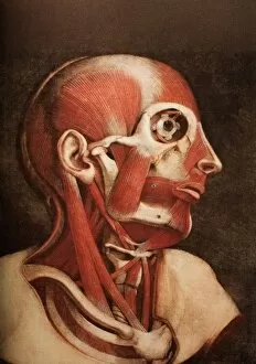
Muscles of the head and neck, historical anatomical artwork. This head has been dissected to show several muscles involved in moving the face and neck

Background imageLarynx Collection: Computer artwork of healthy human lungs

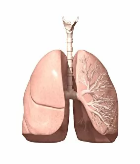
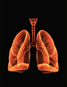
Computer artwork of healthy human lungs

Background imageLarynx Collection: Human respiratory system, artwork

Human respiratory system, artwork
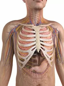
Respiratory system. Computer artwork of a male torso and the respiratory System, showing lungs, bronchus, bronchioles, larynx and windpipe

Background imageLarynx Collection: Thyroid gland, artwork

Thyroid gland, artwork. This two-lobed endocrine gland is found in the neck, in front of the trachea (windpipe, striped). The larynx (voice box, orange) sits above it

Background imageLarynx Collection: Epiglottis, light micrograph

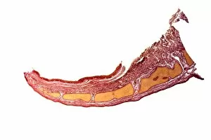
Epiglottis, light micrograph
Epiglottis. Light micrograph of a longitudinal section through the epiglottis, the flap of tissue located at the top of the larynx (voicebox)

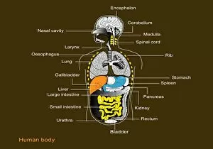
Background imageLarynx Collection: Human internal organs, diagram

Human internal organs, diagram
Human internal organs. Diagram showing the human internal organs in the head and torso. In the head, the organs and cavities shown are: the brain (encephalon), the cerebellum (part of the brain)

Background imageLarynx Collection: Human internal organs, artwork

Human internal organs, artwork
Human internal organs. Artwork showing the human internal organs in the head and torso. In the head, the organs and cavities shown are: the brain (encephalon), the cerebellum (part of the brain)


All Professionally Made to Order for Quick Shipping

The larynx, also known as the voice box, is a vital part of the human respiratory system and can be seen in an illustration depicting the entire respiratory system, including the oral and nasal cavities, trachea, bronchus, and lungs. Another artwork showcases the intricate anatomy of the thyroid gland. Intriguingly, there is an engraving from 1866 that focuses on neck throat anatomy. This piece provides a detailed view of the larynx's position within our bodies. Furthermore, it highlights how this organ plays a crucial role in our ability to speak and produce sound. The importance of the larynx becomes even more apparent when considering its depiction alongside other senses in an artwork titled "Human Senses. " This emphasizes how our vocal abilities are intertwined with our perception of touch, taste, smell, sight, and hearing. Additionally, two artworks show different views of the head from 1746 by Jacques Fabien Gautier Dagoty. These pieces likely include representations of various internal organs such as the larynx. Interestingly enough, advertisements for Geraudels Pastilles may have utilized images or references to promote their product's benefits for soothing throat irritations related to conditions involving the larynx. Medical lithographs provide further insight into specific aspects related to this organ's examination and treatment. One lithograph demonstrates how a laryngoscope can be used to examine vocal apparatuses visually. Another vividly portrays invasive membranes affecting both throat and larynx during diphtheria or croup infections. Finally, a color lithograph shows a polyp being removed from someone's larynx using a similar instrument -the larnygoscope- highlighting medical interventions available for treating certain conditions affecting this delicate structure.