
Jules Germain Cloquet Collection
"Exploring the Intricacies of the Human Body: Jules Germain Cloquet's Artistic Masterpieces" Step into a world where art and anatomy intertwine


All Professionally Made to Order for Quick Shipping
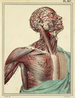
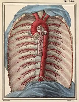
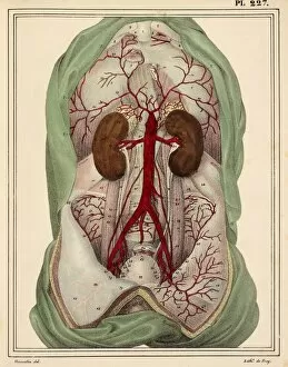
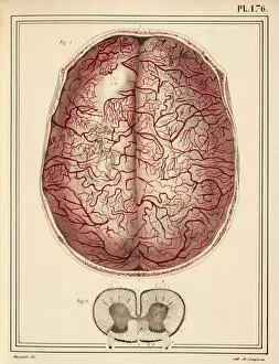
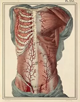
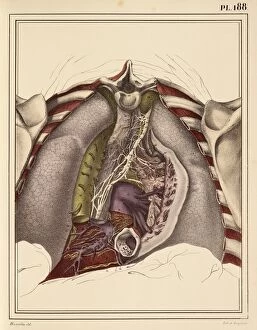
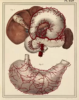
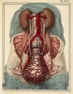
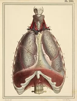
"Exploring the Intricacies of the Human Body: Jules Germain Cloquet's Artistic Masterpieces" Step into a world where art and anatomy intertwine, as we delve into the captivating works of Jules Germain Cloquet. In his remarkable 1825 artwork, he meticulously captures the male groin arteries, revealing the intricate network that sustains life within us. Moving upwards, his attention to detail is evident in his depiction of head and chest arteries. Each stroke on canvas brings forth a mesmerizing display of our vital lifelines pulsating with energy. Cloquet's lithograph from 1865-66 showcases his exceptional talent in portraying human anatomy. His portrayal of face and neck nerves offers an intimate glimpse into our body's complex communication system. Venturing further down, we encounter another masterpiece capturing the thoracic aorta - a testament to Cloquet's dedication to anatomical accuracy. The vibrant colors breathe life into this crucial vessel responsible for carrying oxygen-rich blood throughout our bodies. The artist continues to amaze with his portrayal of intestinal veins, skillfully illustrating their winding paths through delicate brushstrokes. A true visionary ahead of his time. Cloquet's artistic prowess extends beyond organs alone; he also explores liver and stomach nerves in stunning detail. Through these artworks, one can almost feel the intricate connections that govern digestion and metabolism. Intriguingly, Cloquet revisits intestinal veins once again in another artwork from 1825 – perhaps emphasizing their significance or simply captivated by their beauty. A pregnant woman takes center stage in yet another breathtaking creation by Cloquet. With grace and sensitivity, he portrays her form while highlighting the miraculous changes occurring within her body during this transformative period. Abdominal lymph vessels come alive under Cloquet's skilled hand as they weave through tissue with precision and purpose – reminding us how interconnected every part of our being truly is. Delving deeper into human development, Cloquet's artwork unveils the wonders of foetal brain development.