
Ischaemia Collection
"Unveiling the Silent Threat: Exploring Ischaemia through Cutting-Edge Imaging Techniques" Ischaemia, a condition characterized by restricted blood flow to vital organs


All Professionally Made to Order for Quick Shipping
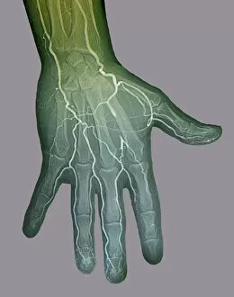
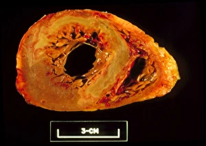
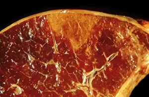
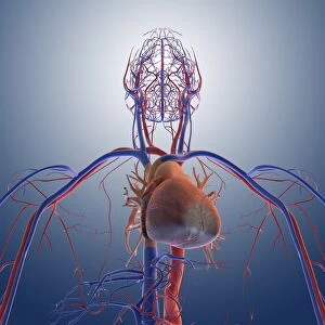
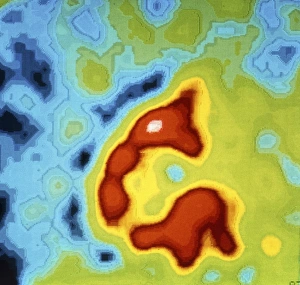
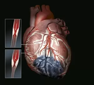
"Unveiling the Silent Threat: Exploring Ischaemia through Cutting-Edge Imaging Techniques" Ischaemia, a condition characterized by restricted blood flow to vital organs, has long been a medical enigma. Thanks to advancements in digital angiogram technology, we can now delve into the intricate world of this silent threat. Stroke, one of the most devastating consequences of ischaemia, comes into focus as MRI and 3D CT scans provide us with unprecedented insights. These powerful imaging tools allow us to visualize the intricate pathways affected by this life-altering event (C016 / 6419). But it's not just strokes that fall under our investigative lens. Splenic infarct, another consequence of ischaemic events, reveals its secrets through detailed imagery (C015 / 6220). Even the ischaemic bowel unveils itself under scrutiny in a captivating light micrograph (C015 / 6219). The human head becomes an open book as MRI and 3D CT scans unravel the mysteries hidden within its complex structure. We witness how ischaemia disrupts these delicate networks and alters lives forever. Atherosclerosis takes center stage in a mesmerizing artwork capturing its destructive impact on arteries (C016 / 6571). Meanwhile, splenic infarcts continue their visual narrative with striking images that showcase their unique characteristics (C015 / 6221). Heart attacks become vivid works of art themselves as we explore them from different angles - each artwork telling a story of pain and resilience (C016 /2895; C016/2894; C016/2893). Scintigrams post-heart attack reveal lingering signs - reminders that even after surviving such an ordeal, challenges persist. Through these groundbreaking imaging techniques, we are beginning to demystify ischaemia's complexities. As science progresses hand-in-hand with technology, we inch closer towards better understanding this silent threat and finding innovative ways to combat its devastating consequences.