Fraud Blocker Skip to main content

Invertebrates Collection (page 11)

"Invertebrates

Background imageInvertebrates Collection: Types of protozoa flagellata: Tripanosoma Abissinicum, belonging to the Bovis species

Types of protozoa flagellata: Tripanosoma Abissinicum, belonging to the Bovis species, enlarged under a microscope. Date of Photograph:1872-1915 ca

Background imageInvertebrates Collection: Mastigophora flagellata: Tripanosoma abissinicum and Piroplasma tigeminum enlarged under a

Mastigophora flagellata: Tripanosoma abissinicum and Piroplasma tigeminum enlarged under a microscope. Date of Photograph:1872-1915 ca

Background imageInvertebrates Collection: Phagocytosis of the protozoa, flagellata: Tripanosoma-Pirosoma, enlarged under a microscope

Phagocytosis of the protozoa, flagellata: Tripanosoma-Pirosoma, enlarged under a microscope. Date of Photograph:1872-1915 ca

Background imageInvertebrates Collection: Diaptomus Castor (Crostacei. Entomostracee) enlarged under a microscope

Diaptomus Castor (Crostacei. Entomostracee) enlarged under a microscope. Date of Photograph:11/1897

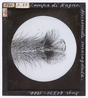
Background imageInvertebrates Collection: Micromata smaragdula spider leg, magnified under a microscope

Micromata smaragdula spider leg, magnified under a microscope. Date of Photograph:19/04/1894

Background imageInvertebrates Collection: Hyperia trigona. (Amphipoda. Hyperina) magnified under the microscope

Hyperia trigona. (Amphipoda. Hyperina) magnified under the microscope. Date of Photograph:04/1914

Background imageInvertebrates Collection: Gammarus pulex (Amphipoda, Gammaridae) magnified under the microscope

Gammarus pulex (Amphipoda, Gammaridae) magnified under the microscope. Date of Photograph:04/1914

Background imageInvertebrates Collection: Corophium longicorne (Amphipoda, Gammaridae) magnified under the microscope

Corophium longicorne (Amphipoda, Gammaridae) magnified under the microscope. Date of Photograph:04/1914

Background imageInvertebrates Collection: Pediculus Capitis (Apteri) enlarged under a microscope

Pediculus Capitis (Apteri) enlarged under a microscope. Date of Photograph:04/1914

Background imageInvertebrates Collection: Phylloxera Vastatrix root louse (Aphidae) enlarged under a microscope

Phylloxera Vastatrix root louse (Aphidae) enlarged under a microscope. Date of Photograph:1914

Background imageInvertebrates Collection: Cornea of the eye of a Tabanus Bovinus (Tabanidae) enlarged under a microscope

Cornea of the eye of a Tabanus Bovinus (Tabanidae) enlarged under a microscope. Date of Photograph:04/1895

Background imageInvertebrates Collection: Haematopota pluvialis (Diptera. Brachycera Fam. Tabanidae), from A. Celli, 'Manuale dell'igienista

Haematopota pluvialis (Diptera. Brachycera Fam. Tabanidae), from A. Celli, "Manuale dell'igienista, ad uso di ufficiali sanitari, medici circondariali e provinciali, ingegneri

Background imageInvertebrates Collection: Cordylobia anthropophaga female and larva (Diptera) from A. Celli, 'Manuale dell'igienista

Cordylobia anthropophaga female and larva (Diptera) from A. Celli, "Manuale dell'igienista, ad uso di ufficiali sanitari, medici circondariali e provinciali, ingegneri

Background imageInvertebrates Collection: Stomoxys calcitrans. (Diptera. Brachycera Muscidae), enlarged under the microscope from A

Stomoxys calcitrans. (Diptera. Brachycera Muscidae), enlarged under the microscope from A. Celli, "Manuale dell'igienista, ad uso di ufficiali sanitari, medici circondariali e provinciali

Background imageInvertebrates Collection: Psycoda phalaenoides (Culiciformes), enlarged under the microscope

Psycoda phalaenoides (Culiciformes), enlarged under the microscope. Date of Photograph:1914

Background imageInvertebrates Collection: Scatophaga leg fly, magnified under the microscope

Scatophaga leg fly, magnified under the microscope. Date of Photograph:19/04/1894

Background imageInvertebrates Collection: Head of male Anopheles, magnified under the microscope

Head of male Anopheles, magnified under the microscope. Date of Photograph:1900-1915 ca

Background imageInvertebrates Collection: Culex pipiens and Anopheles claviger larvae, enlarged under the microscope

Culex pipiens and Anopheles claviger larvae, enlarged under the microscope. Date of Photograph:1890-1914 ca

Background imageInvertebrates Collection: Head of female Anapheles (Diptera) enlarged under a microscope

Head of female Anapheles (Diptera) enlarged under a microscope. Date of Photograph:1872-1915 ca

Background imageInvertebrates Collection: Culex (Diptera) larvae enlarged under a microscope

Culex (Diptera) larvae enlarged under a microscope. Date of Photograph:1872-1915 ca

Background imageInvertebrates Collection: Culex pipiens and Anopheles maculipennis in resting state, fig. 22 from A

Culex pipiens and Anopheles maculipennis in resting state, fig. 22 from A. Celli, Manuale dell'igienista, ad uso di ufficiali sanitari, medici circondariali e provinciali, ingegneri

Background imageInvertebrates Collection: Culex and Anopheles eggs enlarged, fig. 19 from A. Celli, Manuale dell'igienista

Culex and Anopheles eggs enlarged, fig. 19 from A. Celli, Manuale dell'igienista, ad uso di ufficiali sanitari, medici circondariali e provinciali, ingegneri, chimici e veterinari igienisti

Background imageInvertebrates Collection: Dytiscus Natator (Dytiscidae) enlarged under a microscope

Dytiscus Natator (Dytiscidae) enlarged under a microscope. Date of Photograph:1914

Background imageInvertebrates Collection: Pulex canis (Pulicidae) magnified under the microscope

Pulex canis (Pulicidae) magnified under the microscope. Date of Photograph:15/04/1894

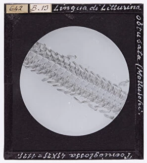
Background imageInvertebrates Collection: Microscopically enlarged tongue of Litturina obtusata (Molluscs)

Microscopically enlarged tongue of Litturina obtusata (Molluscs). Date of Photograph:05/1895

Background imageInvertebrates Collection: Tongue of Helix pomatia (Molluscs) magnified under a microscope

Tongue of Helix pomatia (Molluscs) magnified under a microscope. Date of Photograph:02/1890

Background imageInvertebrates Collection: QFA-F-000138-0000

QFA-F-000138-0000
School of giant medusas in the waters of the Pacific Ocean around the Paracas Peninsula, Per Peru. Date of Photograph:1982

Background imageInvertebrates Collection: A dragonfly and a flying deer, pen drawing on reddish paper by Leonardo da Vinci

A dragonfly and a flying deer, pen drawing on reddish paper by Leonardo da Vinci and preserved at the Royal Library of Turin

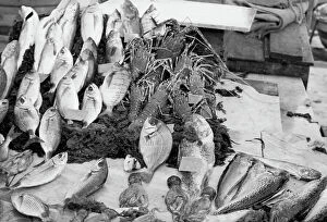
Background imageInvertebrates Collection: School of fish

School of fish Palermo. Date of Photograph:1960-1961

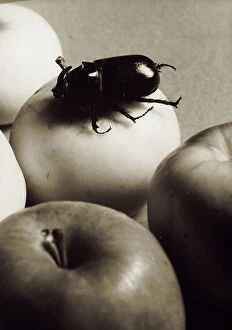
Background imageInvertebrates Collection: A coleoptera on an apple. Postcard sent by the photographer to Vincenzo Balocchi

A coleoptera on an apple. Postcard sent by the photographer to Vincenzo Balocchi Italy. Date of Photograph:1942

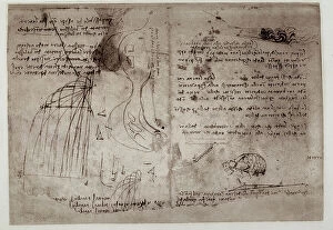
Background imageInvertebrates Collection: Study on waterfalls, written by Leonardo da Vinci, part of the Arundel Codex 263, c.36r

Study on waterfalls, written by Leonardo da Vinci, part of the Arundel Codex 263, c.36r, housed in the British Museum of London Drawing High Renaissance, Renaissance

Background imageInvertebrates Collection: Two crabs; pen drawing on white paper by Leonardo da Vinci preserved in the Wallraff-Richartz

Two crabs; pen drawing on white paper by Leonardo da Vinci preserved in the Wallraff-Richartz Museum in Cologne. Drawing High Renaissance, Renaissance, Renaissance-Baroque styles and periods, Europe

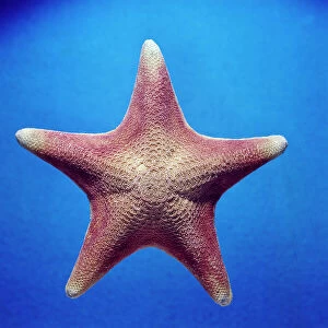
Background imageInvertebrates Collection: Starfish (Patiria Miniata) of California

Starfish (Patiria Miniata) of California. Date of Photograph:1967

Background imageInvertebrates Collection: Helix (Mollusca Gastropoda)

Helix (Mollusca Gastropoda). Date of Photograph:1967

Background imageInvertebrates Collection: Cardium tuberculatum Genoa

Cardium tuberculatum Genoa. Date of Photograph:1967

Background imageInvertebrates Collection: Acauus haemastoma Sri Lanka

Acauus haemastoma Sri Lanka Sri Lanka ( Ceylon ). Date of Photograph:1967

Background imageInvertebrates Collection: Octopus (Octopus), Civic Aquarium of Milan

Octopus (Octopus), Civic Aquarium of Milan Milan Civic Aquarium of Milan. Date of Photograph:1967

Background imageInvertebrates Collection: Julidae (Diplopoda)

Julidae (Diplopoda) Africa. Date of Photograph:1967

Background imageInvertebrates Collection: Marine copepod eggs

Marine copepod eggs Africa. Date of Photograph:1967

Background imageInvertebrates Collection: Marine planarian (Prostheceracus), Gulf of Naples

Marine planarian (Prostheceracus), Gulf of Naples Naples. Date of Photograph:1967

Background imageInvertebrates Collection: Still life with flowers, fruit and insects; painting by Rachel Ruysch

Still life with flowers, fruit and insects; painting by Rachel Ruysch. Galleria Palatina, Palazzo Pitti, Florence Painting Eighteenth Century, Renaissance-Baroque styles and periods, Europe

Background imageInvertebrates Collection: Birth of Venus; work of Sandro Botticelli. Uffizi Gallery, Florence

Birth of Venus; work of Sandro Botticelli. Uffizi Gallery, Florence Painting Early Renaissance, Renaissance, Renaissance-Baroque styles and periods, Europe, First and Second Millennium A.D

Background imageInvertebrates Collection: Helix (Gasteropoda mollusca)

Helix (Gasteropoda mollusca). Date of Photograph:1967

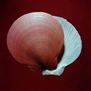
Background imageInvertebrates Collection: Amussium Pleuronectes Indian Ocean

Amussium Pleuronectes Indian Ocean. Date of Photograph:1967

Background imageInvertebrates Collection: Cypraea tigris Pacific Ocean

Cypraea tigris Pacific Ocean. Date of Photograph:1967

Background imageInvertebrates Collection: Head Sagitta (Chaetognatha Aphragmorpha Sagittoidei)

Head Sagitta (Chaetognatha Aphragmorpha Sagittoidei) Africa. Date of Photograph:1967

Background imageInvertebrates Collection: A jellyfish (Pelagia Noctiluca)

A jellyfish (Pelagia Noctiluca) Italy. Date of Photograph:01-03/1968

Background imageInvertebrates Collection: Birth of Venus, detail, tempera on canvas, Botticelli, Sandro (1445-1510), Uffizi Gallery, Florence

Birth of Venus, detail, tempera on canvas, Botticelli, Sandro (1445-1510), Uffizi Gallery, Florence Florence Uffizi Gallery Painting Early Renaissance, Renaissance


All Professionally Made to Order for Quick Shipping

"Invertebrates: A Fascinating World of Diversity and Adaptation" From the intricate honeycombs built by diligent honey bees to the captivating sight of a lobster's inner workings revealed through an X-ray, invertebrates never cease to amaze us. The Red Admiral butterfly gracefully displays its vibrant wings, showcasing nature's artistic brilliance. Meanwhile, under the lens of a scanning electron microscope, calcareous phytoplankton reveal their delicate beauty as they float effortlessly in the vast ocean. In Kagoshima Prefecture, Kyushu, Japan, a male Leach's sea star steals the spotlight as it engages in broadcast spawning - an awe-inspiring phenomenon where streams of sperm are released from its arms into the water. This mesmerizing moment captured by a talented wildlife photographer earned them recognition as the winner in the Underwater category at Wildlife Photographer of the Year 2022. Venturing into Croatian waters along Korcula Island in the Adriatic Sea reveals another enchanting creature - a Nudibranch known as Janolus cristatus. Its vibrant colors and graceful movements make it a true gem beneath the waves. A sea green swallowtail butterfly flutters elegantly across meadows and gardens while fruit flies unveil their intricate structures when observed under high-resolution SEM imaging techniques. Delving deep into Earth's history brings us face-to-face with ancient wonders like trilobite fossils that offer glimpses into prehistoric times. These extinct arthropods remind us of our planet's rich evolutionary past. Witnessing an ocellate octopus swimming up from the depths is like witnessing magic unfold before our eyes. Its ethereal appearance leaves us captivated by nature's ability to create such extraordinary creatures. Lastly, we encounter a painted lady butterfly whose delicate patterns adorn fields with splashes of color and gracefulness. And even within microscopic realms lies astonishing complexity; for instance, observing dog tapeworm heads through SEM reveals the intricate structures that allow these parasites to thrive.