Fraud Blocker Skip to main content

Invertebrates Collection (page 9)

"Invertebrates

Background imageInvertebrates Collection: Postcard depicting a woman in a forest with a butterfly, 'Album para Tarjetas postales'

Postcard depicting a woman in a forest with a butterfly, "Album para Tarjetas postales" Europe. Date of Photograph:1910 ca

Background imageInvertebrates Collection: An oyster (ostrea edulis) in a rocky sea depth

An oyster (ostrea edulis) in a rocky sea depth. Europe. Date of Photograph:1960-1965 ca

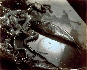
Background imageInvertebrates Collection: Still life with fish and polyps

Still life with fish and polyps. Date of Photograph:1934

Background imageInvertebrates Collection: Still life with polyps and fish

Still life with polyps and fish. Date of Photograph:1934

Background imageInvertebrates Collection: Pictorialist still life with fish and polyps

Pictorialist still life with fish and polyps. Date of Photograph:1934

Background imageInvertebrates Collection: Two young Japanese women in traditional costume during the processing of silkworms

Two young Japanese women in traditional costume during the processing of silkworms Japan. Date of Photograph:1885-1895

Background imageInvertebrates Collection: Venetian costumes and masks: peddler of mussels and oysters

Venetian costumes and masks: peddler of mussels and oysters Venice. Date of Photograph:1860 ca

Background imageInvertebrates Collection: Rearing of silkworms for the textile industry, Japan

Rearing of silkworms for the textile industry, Japan Japan. Date of Photograph:1890-1899

Background imageInvertebrates Collection: Album 'Von Herr August Egli in Jokohama - Juni 1895': two young Japanese women in traditional

Album "Von Herr August Egli in Jokohama - Juni 1895": two young Japanese women in traditional costume settle silkworms useful to the textile industry Japan Yokohama

Background imageInvertebrates Collection: Two butterflies on the leaf of a plant

Two butterflies on the leaf of a plant. Europe. Date of Photograph:1955 - 1960

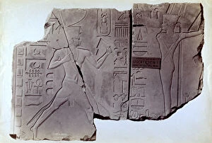
Background imageInvertebrates Collection: Fragment of an Egyptian bas-relief from the Temple of Usertesen I, in the British Museum in London

Fragment of an Egyptian bas-relief from the Temple of Usertesen I, in the British Museum in London Great Britain London, British Museum Low Relief, Basrelief Egypt - Egyptian Art, Africa

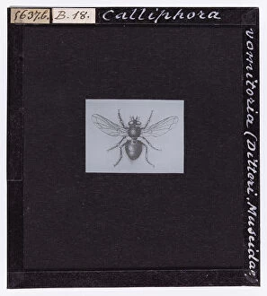
Background imageInvertebrates Collection: Calliphora Vomitoria (Diptera. Muscidae), from A. Celli, 'Manuale dell'igienista

Calliphora Vomitoria (Diptera. Muscidae), from A. Celli, "Manuale dell'igienista, ad uso di ufficiali sanitari, medici circondariali e provinciali, ingegneri, chimici e veterinari igienisti

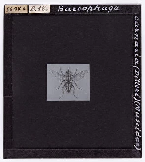
Background imageInvertebrates Collection: Sarcophaga carnaria (Diptera)(Muscidae) from A. Celli, 'Manuale dell'igienista

Sarcophaga carnaria (Diptera)(Muscidae) from A. Celli, "Manuale dell'igienista, ad uso di ufficiali sanitari, medici circondariali e provinciali, ingegneri, chimici e veterinari igienisti

Background imageInvertebrates Collection: Female Anopheles simplex, magnified under the microscope

Female Anopheles simplex, magnified under the microscope. Date of Photograph:1890-1914 ca

Background imageInvertebrates Collection: Male Anopheles simplex, magnified under the microscope

Male Anopheles simplex, magnified under the microscope. Date of Photograph:1890-1914 ca

Background imageInvertebrates Collection: Female Anopheles maculiformis, magnified under the microscope

Female Anopheles maculiformis, magnified under the microscope. Date of Photograph:1890-1914 ca

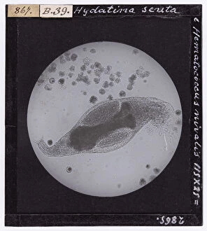
Background imageInvertebrates Collection: Hydatina scrita and Hematococcus nivalis protozoa enlarged under a microscope

Hydatina scrita and Hematococcus nivalis protozoa enlarged under a microscope. Date of Photograph:1913

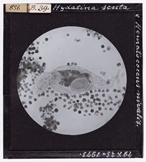
Background imageInvertebrates Collection: Hydatina scrita and Hematacoccus nivalis micro-organisms, enlarged under a microscope

Hydatina scrita and Hematacoccus nivalis micro-organisms, enlarged under a microscope. Date of Photograph:04/1913

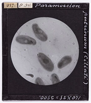
Background imageInvertebrates Collection: Ciliate protozoa, Paramecium putrinum, enlarged under a microscope

Ciliate protozoa, Paramecium putrinum, enlarged under a microscope. Date of Photograph:04/1914

Background imageInvertebrates Collection: Carchesium polypinum: ciliate protozoa enlarged under a microscope

Carchesium polypinum: ciliate protozoa enlarged under a microscope. Date of Photograph:1897

Background imageInvertebrates Collection: Negri bodies present in the cell of the of the Horn of Ammon of a rabid cow

Negri bodies present in the cell of the of the Horn of Ammon of a rabid cow, enlarged under a microscope. Date of Photograph:1900-1915 ca

Background imageInvertebrates Collection: Protozoa of babesia canis in bone marrow: blood parasite in mammals

Protozoa of babesia canis in bone marrow: blood parasite in mammals, enlarged under a microscope (Haemosproidia). Date of Photograph:1872-1915 ca

Background imageInvertebrates Collection: Babesia canis in bone marrow, enlarged under a microscope

Babesia canis in bone marrow, enlarged under a microscope. Date of Photograph:1872-1915 ca

Background imageInvertebrates Collection: Plasmodium of the Colobus Guereza monkey, enlarged under a microscope

Plasmodium of the Colobus Guereza monkey, enlarged under a microscope. Date of Photograph:1872-1915 ca

Background imageInvertebrates Collection: Microscopic enlargement of the parasite of the Tertian fever

Microscopic enlargement of the parasite of the Tertian fever. Date of Photograph:1900-1915 ca

Background imageInvertebrates Collection: Microscopic enlargment of the malaria cists in the stomach of Anopheles, the malaria mosquito

Microscopic enlargment of the malaria cists in the stomach of Anopheles, the malaria mosquito. Date of Photograph:1900-1915 ca

Background imageInvertebrates Collection: Miocroscopic enlargement of Sporozoi, parasite protozoans

Miocroscopic enlargement of Sporozoi, parasite protozoans. Date of Photograph:1888-1915 ca

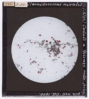
Background imageInvertebrates Collection: Clamidococcus nivalis (Alga) enlarged under a microscope. The protozoa fell with the rain in

Clamidococcus nivalis (Alga) enlarged under a microscope. The protozoa fell with the rain in October 1900, in Florence. Date of Photograph:12/1902

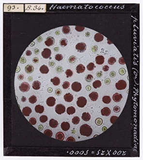
Background imageInvertebrates Collection: Haematococcus pluviatis: Phitomonadino, enlarged under a microscope

Haematococcus pluviatis: Phitomonadino, enlarged under a microscope. Date of Photograph:1888

Background imageInvertebrates Collection: Section of the stomach of an Anopheles, Amfionte (Zygote); fertilized macrospore in this image

Section of the stomach of an Anopheles, Amfionte (Zygote); fertilized macrospore in this image, enlarged under a microscope. Date of Photograph:1888

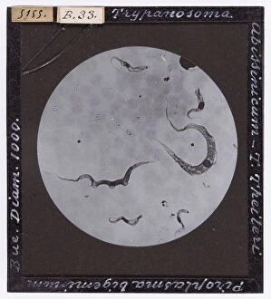
Background imageInvertebrates Collection: Types of Tripanosoma Abissinicum, Theileri, and Piroplasma ligem, belonging to the Bovis species

Types of Tripanosoma Abissinicum, Theileri, and Piroplasma ligem, belonging to the Bovis species, enlarged under a microscope. Date of Photograph:1872-1915 ca

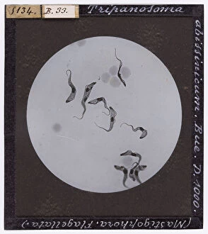
Background imageInvertebrates Collection: Protozoa flagellata: Tripanosoma abissinicum found in the ox, enlarged under a microscope

Protozoa flagellata: Tripanosoma abissinicum found in the ox, enlarged under a microscope. Date of Photograph:1872-1915 ca

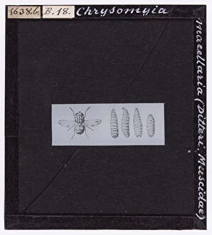
Background imageInvertebrates Collection: Chrysomyia macellaria (Diptera. Muscidae) from A. Celli, 'Manuale dell'igienista

Chrysomyia macellaria (Diptera. Muscidae) from A. Celli, "Manuale dell'igienista, ad uso di ufficiali sanitari, medici circondariali e provinciali, ingegneri, chimici e veterinari igienisti

Background imageInvertebrates Collection: Female Anopheles punctatus, magnified under the microscope

Female Anopheles punctatus, magnified under the microscope. Date of Photograph:1890-1914 ca

Background imageInvertebrates Collection: Spirochaeta pallida bacterium: unicellular micro-organism with a stretched spiral shape

Spirochaeta pallida bacterium: unicellular micro-organism with a stretched spiral shape, enlarged under a microscope. Date of Photograph:1900-1915 ca

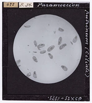
Background imageInvertebrates Collection: Paramoecion putrinum: ciliate protozoa enlarged under a microscope

Paramoecion putrinum: ciliate protozoa enlarged under a microscope. Date of Photograph:1897

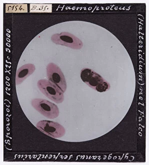
Background imageInvertebrates Collection: Bigeminum babesia in oxen: protozoa enlarged under a microscope(Haemosproidia)

Bigeminum babesia in oxen: protozoa enlarged under a microscope(Haemosproidia). Date of Photograph:1872-1915 ca

Background imageInvertebrates Collection: Tropical babesia in oxen: illness owed to parasite protozoa (pyroplasma) in the blood of mammals

Tropical babesia in oxen: illness owed to parasite protozoa (pyroplasma) in the blood of mammals, transmitted by ticks (Haemosproidia). Enlarged under a microscope. Date of Photograph:1872-1915 ca

Background imageInvertebrates Collection: Pigeon blood with Datulewski Plasmodium, enlarged under a microscope

Pigeon blood with Datulewski Plasmodium, enlarged under a microscope. Date of Photograph:1900-1915 ca

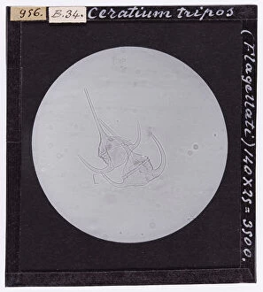
Background imageInvertebrates Collection: Ceratium tripos: protozoa belonging to the Flagellati, enlarged under a microscope

Ceratium tripos: protozoa belonging to the Flagellati, enlarged under a microscope. Date of Photograph:04/1914

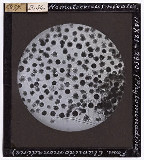
Background imageInvertebrates Collection: Hematococcus nivalis, belonging to the Clamido-monadine family, enlarged under a microscope

Hematococcus nivalis, belonging to the Clamido-monadine family, enlarged under a microscope. Date of Photograph:04/1913

Background imageInvertebrates Collection: Tripanosoma in the blood, enlarged under a microscope

Tripanosoma in the blood, enlarged under a microscope. Date of Photograph:1900-1915 ca

Background imageInvertebrates Collection: Protozoa flagellata: Tripanosoma abissinicum found in horses, enlarged under a microscope

Protozoa flagellata: Tripanosoma abissinicum found in horses, enlarged under a microscope. Date of Photograph:1872-1915 ca

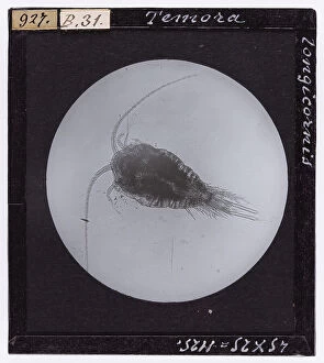
Background imageInvertebrates Collection: Microscopic enlargment of Temora Longicornis

Microscopic enlargment of Temora Longicornis. Date of Photograph:1914

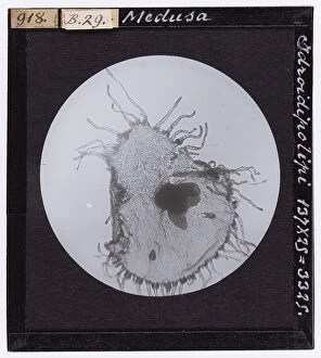
Background imageInvertebrates Collection: Jellyfish (Hydroid polyp) enlarged under a microscope

Jellyfish (Hydroid polyp) enlarged under a microscope. Date of Photograph:1914

Background imageInvertebrates Collection: Agelena spider leg enlarged under a microscope

Agelena spider leg enlarged under a microscope. Date of Photograph:19/04/1894

Background imageInvertebrates Collection: Phylloxera Vastatrix (Aphida. Emiptera) enlarged under a microscope

Phylloxera Vastatrix (Aphida. Emiptera) enlarged under a microscope. Date of Photograph:1914

Background imageInvertebrates Collection: Sting and proboscis of Tabanus bovinus (Tabanidae) magnified under a microscope

Sting and proboscis of Tabanus bovinus (Tabanidae) magnified under a microscope. Date of Photograph:15/04/1894


All Professionally Made to Order for Quick Shipping

"Invertebrates: A Fascinating World of Diversity and Adaptation" From the intricate honeycombs built by diligent honey bees to the captivating sight of a lobster's inner workings revealed through an X-ray, invertebrates never cease to amaze us. The Red Admiral butterfly gracefully displays its vibrant wings, showcasing nature's artistic brilliance. Meanwhile, under the lens of a scanning electron microscope, calcareous phytoplankton reveal their delicate beauty as they float effortlessly in the vast ocean. In Kagoshima Prefecture, Kyushu, Japan, a male Leach's sea star steals the spotlight as it engages in broadcast spawning - an awe-inspiring phenomenon where streams of sperm are released from its arms into the water. This mesmerizing moment captured by a talented wildlife photographer earned them recognition as the winner in the Underwater category at Wildlife Photographer of the Year 2022. Venturing into Croatian waters along Korcula Island in the Adriatic Sea reveals another enchanting creature - a Nudibranch known as Janolus cristatus. Its vibrant colors and graceful movements make it a true gem beneath the waves. A sea green swallowtail butterfly flutters elegantly across meadows and gardens while fruit flies unveil their intricate structures when observed under high-resolution SEM imaging techniques. Delving deep into Earth's history brings us face-to-face with ancient wonders like trilobite fossils that offer glimpses into prehistoric times. These extinct arthropods remind us of our planet's rich evolutionary past. Witnessing an ocellate octopus swimming up from the depths is like witnessing magic unfold before our eyes. Its ethereal appearance leaves us captivated by nature's ability to create such extraordinary creatures. Lastly, we encounter a painted lady butterfly whose delicate patterns adorn fields with splashes of color and gracefulness. And even within microscopic realms lies astonishing complexity; for instance, observing dog tapeworm heads through SEM reveals the intricate structures that allow these parasites to thrive.