
Human Immunodeficiency Virus Collection
"HIV: Unveiling the Silent Intruder" In a world filled with microscopic battles, the HIV particle emerges as a formidable foe


All Professionally Made to Order for Quick Shipping
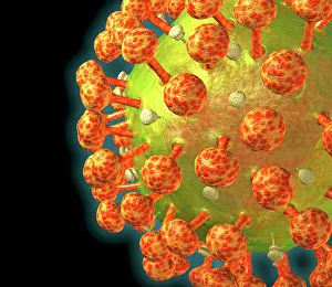
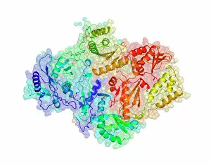
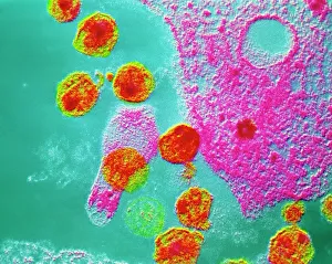
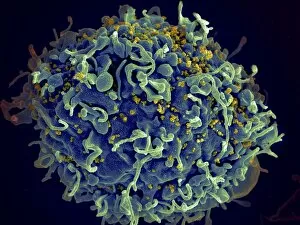
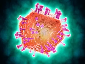
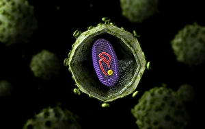
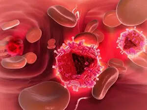
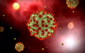
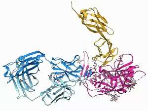
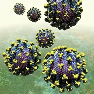
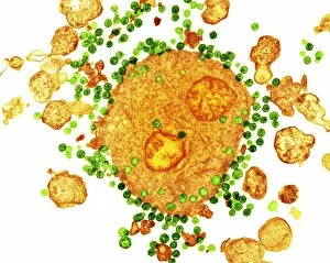
"HIV: Unveiling the Silent Intruder" In a world filled with microscopic battles, the HIV particle emerges as a formidable foe. Its deceptive simplicity belies its destructive power, as it infiltrates human cells and hijacks their machinery. The HIV reverse transcription enzyme cunningly converts its genetic material into DNA, seamlessly integrating itself into our very essence. Through the lens of science, we witness a false-colored TEM image capturing the AIDS virus lurking within a T-cell—a chilling reminder of the relentless assault on our immune system. As if mocking our defenses, AIDS viruses bud from infected cells in another TEM snapshot—an eerie dance of life and death. Amidst this medical turmoil, actor Douglas Lambert bravely faces his diagnosis at St Stephens Hospital. His journey becomes an emblematic representation of countless lives affected by this devastating disease. In North London, Doug's home transforms into both sanctuary and battleground as he valiantly fights against AIDS' merciless grip. The 19th of January marks an important milestone—the opening day for Middlesex Hospital's Broderip AIDS ward—a beacon of hope amidst despair. This dedicated space becomes a haven for those battling against this insidious virus; where compassion meets expertise to provide solace in times of anguish. Time moves forward relentlessly but not without leaving indelible imprints on humanity's fight against HIV/AIDS. On October 31st, 1986—on that fateful Halloween night—Doug succumbs to his battle with AIDS; yet his legacy endures through awareness and advocacy efforts that continue to shape society. A year later, December 16th witnesses another chapter unfold at Middlesex Hospital—the Broderip AIDS ward stands resolute in its mission to care for those afflicted by this epidemic scourge. It serves as a testament to resilience and determination in combating an adversary that knows no boundaries.