Fraud Blocker Skip to main content

Hormonal Collection

"Hormonal: Unveiling the Intricacies of our Body's Chemical Messengers" Delving into the depths of our brain, the medulla oblongata holds the key to hormonal regulation

Background imageHormonal Collection: Medulla oblongata in the brain, artwork

Medulla oblongata in the brain, artwork
Hypothalamus in the brain. Computer artwork of a persons head showing the brain inside. The highlighted area shows the hypothalamus

Background imageHormonal Collection: Cortisol crystals, light micrograph

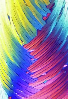
Cortisol crystals, light micrograph
Cortisol crystals, polarised light micrograph. Cortisol is a steroid hormone produced by the adrenal glands, which sit on top of the kidneys

Background imageHormonal Collection: Pancreas anatomy, artwork

Pancreas anatomy, artwork
Pancreas anatomy. Cut-away computer artwork of the pancreas, which secretes digestive enzymes and the hormones insulin and glucagon, which regulate blood sugar levels

Background imageHormonal Collection: Islet of Langerhans, light micrograph

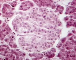
Islet of Langerhans, light micrograph
Islet of Langerhans. Light micrograph of a section through an islet of Langerhans (centre) in the pancreas. This structure is composed of clumps of secretory cells

Background imageHormonal Collection: Oxytocin neurotransmitter molecule

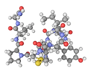
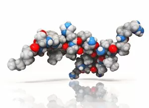
Oxytocin neurotransmitter molecule. Computer model showing the structure of the neurotransmitter and hormone Oxytocin. Atoms are colour-coded spheres (carbon: dark grey, hydrogen: light grey)

Background imageHormonal Collection: Thyroid anatomy, artwork C013 / 4675

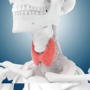
Thyroid anatomy, artwork C013 / 4675
Thyroid anatomy. Computer artwork of the thyroid glands (pink) of the neck, shown in relation to the bodys bones and throat structures (white)

Background imageHormonal Collection: Fight-or-flight response, artwork

Fight-or-flight response, artwork
Fight-or-flight response. Artwork showing the bodys fight-or-flight response to a threat. Visual perception (yellow arrow) can trigger fear memories controlled by the brains amygdala (yellow)

Background imageHormonal Collection: Menopause, conceptual artwork

Menopause, conceptual artwork. Menopause is the cessation of menstruation. It usually occurs between the ages of 45 and 55

Background imageHormonal Collection: Parathyroid hormone molecule

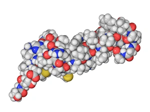
Parathyroid hormone molecule. Computer model showing the structure of parathyroid hormone (PTH), or parathormone. Atoms are colour-coded (carbon: dark grey, hydrogen: light grey, oxygen: red)

Background imageHormonal Collection: Ghrelin hormone molecule

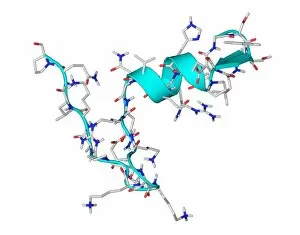
Ghrelin hormone molecule. Computer model showing the crystal structure of the human hormone ghrelin. The crystal structure consists of both the secondary structure

Background imageHormonal Collection: Contraceptive implant, X-ray C017 / 7390

Contraceptive implant, X-ray C017 / 7390
Contraceptive implant. X-ray of the arm of a 19 year old female showing a contraceptive implant (thin rod at upper centre)

Background imageHormonal Collection: Hydrocortisone ointment F008 / 3400

Hydrocortisone ointment F008 / 3400
Hydrocortisone ointment in tube

Background imageHormonal Collection: Psychic brain, conceptual image

Psychic brain, conceptual image. Artwork showing the human brain and head, surrounded in flames, with the pineal gland highlighted (bright dot in centre of forehead)

Background imageHormonal Collection: Insulin molecule F006 / 9761

Insulin molecule F006 / 9761
Insulin molecule. Molecular model of the hormone insulin from a pig. Insulin consists of two peptide chains, A and B, which are linked by disulphide bridges

Background imageHormonal Collection: Insulin molecule F006 / 9625

Insulin molecule F006 / 9625
Insulin, molecular model. Insulin plays an important role in blood sugar regulation. It is released from the pancreas when blood sugar levels are high, for example after a meal

Background imageHormonal Collection: Insulin molecule F006 / 9605

Insulin molecule F006 / 9605
Insulin molecule. Molecular model of the hormone insulin. Insulin consists of two peptide chains, A and B, which are linked by disulphide bridges

Background imageHormonal Collection: Leptin molecule F006 / 9243

Leptin molecule F006 / 9243
Leptin molecule. Molecular model of the human hormone leptin. Leptin is a protein produced by adipose (fat) tissue. It interacts with receptors in the brains hypothalamus to signal when a person is

Background imageHormonal Collection: Kidneys, anatomical artwork

Kidneys, anatomical artwork
Kidneys. Anatomical computer artwork of a mans body, showing the position of the kidneys (red, centre left and right) and adrenal glands (beige)

Background imageHormonal Collection: Renal blood supply, anatomical artwork

Renal blood supply, anatomical artwork
Renal blood supply. Anatomical computer artwork of a human kidney (left) and adrenal gland (beige, upper centre), showing their main blood supply

Background imageHormonal Collection: Diabetic woman, artwork C016 / 9367

Diabetic woman, artwork C016 / 9367
Diabetic woman. Computer artwork showing sugar (white dots) entering the blood stream of a diabetic woman as she eats an apple. Some of the sugar is used by mitochondria (blue) to produce energy

Background imageHormonal Collection: Jealousy research, MRI brain scans C016 / 9633

Jealousy research, MRI brain scans C016 / 9633
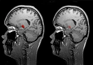
Jealousy research. Magnetic resonance imaging (MRI) brain scans carried out as part of research into human jealousy. The research is being carried out by neuropsychologist Kenneth Levy at

Background imageHormonal Collection: Jealousy, conceptual MRI brain scans C016 / 9634

Jealousy, conceptual MRI brain scans C016 / 9634
Jealousy. Conceptual composite magnetic resonance imaging (MRI) brain scans demonstrating differences in jealousy between the sexes

Background imageHormonal Collection: Corpus luteum, TEM

Corpus luteum, TEM
Corpus luteum. Transmission electron micrograph (TEM) of a section through the corpus luteum of an ovary, showing several luteal cells

Background imageHormonal Collection: Islet of Langerhans beta cell, TEM

Islet of Langerhans beta cell, TEM
Islet of Langerhans beta cell. Transmission electron micrograph (TEM) of a section through a beta cell in an islet of Langerhans in the pancreas

Background imageHormonal Collection: Diabetes, artwork

Diabetes, artwork
Diabetes. Computer artwork showing the difference between a diabetic (right) and non-diabetic (left) persons blood glucose uptake

Background imageHormonal Collection: Cushings syndrome, light micrograph

Cushings syndrome, light micrograph
Cushings syndrome. Light micrograph of a section through muscle affected by Cushings syndrome, a hormonal condition caused by overproduction of adrenal gland hormones

Background imageHormonal Collection: Steroid secreting cell, TEM

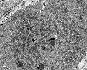
Steroid secreting cell, TEM
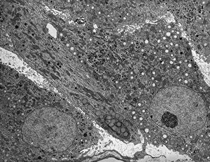
Steroid secreting cell. Transmission electron micrograph (TEM) of a section through a cell involved with steroid synthesis and secretion (steroidogenesis)

Background imageHormonal Collection: Islet of Langerhans, TEM

Islet of Langerhans, TEM
Islet of Langerhans. Transmission electron micrograph (TEM) of a section through cells clustered in an islet of Langerhans in the pancreas

Background imageHormonal Collection: Anterior pituitary gland, TEM

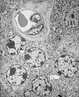
Anterior pituitary gland, TEM
Anterior pituitary gland. Transmission electron micrograph (TEM) of a section through cells of the anterior pituitary. The anterior pituitary, or adenohypophysis

Background imageHormonal Collection: Thyroid follicles, light micrograph

Thyroid follicles, light micrograph
Thyroid follicles. Light micrograph of a section through a thyroid gland showing the follicles. The follicles are lined by a single layer of cuboidal epithelial cells

Background imageHormonal Collection: Enteroendocrine cell, TEM

Enteroendocrine cell, TEM
Enteroendocrine cell. Transmission electron micrograph (TEM) of a section through an enteroendocrine cell within a gland of the stomach

Background imageHormonal Collection: Islet of Langerhans, light micrograph C016 / 0514

Islet of Langerhans, light micrograph C016 / 0514
Islet of Langerhans. Light micrograph of a section through an islet of Langerhans in a pancreas. Scattered throughout the pancreas, the islets are endocrine cells that secrete a range of hormones

Background imageHormonal Collection: Islet of Langerhans, light micrograph C016 / 0513

Islet of Langerhans, light micrograph C016 / 0513
Islet of Langerhans. Light micrograph of a section through an islet of Langerhans in a pancreas. Scattered throughout the pancreas, the islets are endocrine cells that secrete a range of hormones

Background imageHormonal Collection: Psychic brain, conceptual image C014 / 1480

Psychic brain, conceptual image C014 / 1480
Psychic brain, conceptual image. Artwork showing the human brain from the side, surrounded by flames, with the pineal gland shown

Background imageHormonal Collection: Leptin molecule C014 / 4904

Leptin molecule C014 / 4904
Leptin molecule. Computer model showing the structure of a molecule of the hormone leptin. Leptin is produced by adipose (fat) tissue

Background imageHormonal Collection: Peptide YY obesity hormone molecule C014 / 4911

Peptide YY obesity hormone molecule C014 / 4911
Peptide YY obesity hormone molecule. Computer model showing the crystal structure of a molecule of the hormone peptide YY (PYY, or PYY3-36)

Background imageHormonal Collection: Peptide YY obesity hormone molecule C014 / 4910

Peptide YY obesity hormone molecule C014 / 4910
Peptide YY obesity hormone molecule. Computer model showing the crystal structure of a molecule of the hormone peptide YY (PYY, or PYY3-36)

Background imageHormonal Collection: Leptin molecule C014 / 4907

Leptin molecule C014 / 4907
Leptin molecule. Computer model showing the crystal structure of a molecule of the hormone leptin. Leptin is produced by adipose (fat) tissue

Background imageHormonal Collection: Cholecystokinin-8 molecule C014 / 4895

Cholecystokinin-8 molecule C014 / 4895
Cholecystokinin-8 molecule. Computer model showing the structure of the terminal fragment of a molecule of the hormone cholecystokinin-8 (CCK-8)

Background imageHormonal Collection: Activated ghrelin hormone molecule C014 / 4902

Activated ghrelin hormone molecule C014 / 4902
Activated ghrelin hormone molecule. Computer model showing the crystal structure of the human hormone ghrelin. The crystal structure consists of both the secondary structure

Background imageHormonal Collection: Activated ghrelin hormone molecule C014 / 4903

Activated ghrelin hormone molecule C014 / 4903
Activated ghrelin hormone molecule. Computer model showing the structure of the human hormone ghrelin. Atoms are colour-coded spheres (carbon: grey, oxygen: red, nitrogen: blue)

Background imageHormonal Collection: Leptin molecule and fat cells C014 / 4906

Leptin molecule and fat cells C014 / 4906
Leptin molecule and fat cells. Computer artwork showing a molecule of the hormone leptin with adipose (fat) cells (round), from which it is produced

Background imageHormonal Collection: Obestatin molecule C014 / 4908

Obestatin molecule C014 / 4908
Obestatin molecule. Computer artwork showing the structure of a molecule of obestatin. Obestatin is thought to supress hunger and reduce food intake, thereby reducing weight gain

Background imageHormonal Collection: Obestatin molecule C014 / 4909

Obestatin molecule C014 / 4909
Obestatin molecule. Computer artwork showing the structure of a molecule of obestatin. Obestatin is thought to supress hunger and reduce food intake, thereby reducing weight gain

Background imageHormonal Collection: Cholecystokinin-8 molecule C014 / 4894

Cholecystokinin-8 molecule C014 / 4894
Cholecystokinin-8 molecule. Computer model showing the structure of the terminal fragment of a molecule of the hormone cholecystokinin-8 (CCK-8)

Background imageHormonal Collection: Liver, gallbladder and pancreas, artwork

Liver, gallbladder and pancreas, artwork
Liver, gallbladder and pancreas, computer artwork. The largest organ seen here is the liver. Below the liver are the pancreas (pink, left to right) and the gallbladder (brown, left)

Background imageHormonal Collection: Activated ghrelin hormone molecule C014 / 4901

Activated ghrelin hormone molecule C014 / 4901
Activated ghrelin hormone molecule. Computer model showing the crystal structure of the human hormone ghrelin. The crystal structure consists of both the secondary structure

Background imageHormonal Collection: Vasopressin hormone crystals, LM C016 / 8551

Vasopressin hormone crystals, LM C016 / 8551
Vasopressin hormone crystals, light micrograph taken with polarised light. Vasopressin is a neurohypophysial hormone found in most mammals


All Professionally Made to Order for Quick Shipping

"Hormonal: Unveiling the Intricacies of our Body's Chemical Messengers" Delving into the depths of our brain, the medulla oblongata holds the key to hormonal regulation. Behold the mesmerizing artwork capturing cortisol crystals under a light micrograph - a glimpse into stress management at its core. Explore the intricate anatomy of the pancreas through captivating artwork, where hormones play their symphony. Witness the beauty within Islets of Langerhans under a light micrograph, revealing their crucial role in blood sugar control. The oxytocin neurotransmitter molecule - an enchanting messenger that fosters love and bonding in our brains. Dive into an artistic representation of thyroid anatomy, unraveling its significance in metabolism and energy balance. Marvel at SEM images showcasing thyroid gland capillaries and blood vessels - lifelines for hormone distribution throughout our body. Journey through the brain's limbic system, where emotions intertwine with hormonal signals to shape our experiences. Admire exquisite artwork depicting Islets of Langerhans cells - guardians ensuring harmony within our endocrine system. Embark on an anatomical exploration of kidney function through stunning artwork, highlighting its role in hormone production and filtration processes. In this captivating journey, we uncover how these hormonal cues orchestrate countless bodily functions with precision and grace – truly a testament to nature's brilliance.