Fraud Blocker Skip to main content

Hepatocyte Collection

A hepatocyte, also known as a liver cell, is a vital component of the liver tissue

Background imageHepatocyte Collection: Hepatocyte liver cell, TEM

Hepatocyte liver cell, TEM
Hepatocyte liver cell. Transmission electron micrograph (TEM) of a section through an hepatocyte liver cell, showing rough and smooth endoplasmic reticulum (ER, dark lines)

Background imageHepatocyte Collection: Liver tissue, TEM

Liver tissue, TEM
Liver tissue. Transmission electron micrograph (TEM) of a section through the liver, showing part of a radial cord of hepatocyte liver cells (dark) and the vascular sinusoids (white)

Background imageHepatocyte Collection: Laminae formed by hepatocytes

Laminae formed by hepatocytes
Medicine - Human Histology - Digestive system - Liver - Hepatic tissue - Laminae formed by hepatocytes under the microscope

Background imageHepatocyte Collection: Cirrhosis of the liver, light micrograph

Cirrhosis of the liver, light micrograph
Cirrhosis of the liver. Light micrograph of a section through liver tissue affected by cirrhosis, showing a cluster of hepatocyte cells (orange)

Background imageHepatocyte Collection: Cirrhosis of liver, light micrograph C016 / 0530

Cirrhosis of liver, light micrograph C016 / 0530
Cirrhosis of liver. Light micrograph of a section through liver tissue, damaged by cirrhosis. Cirrhosis occurs as a result of a range of factors causing damage to liver function

Background imageHepatocyte Collection: Cirrhosis of liver, light micrograph C016 / 0529

Cirrhosis of liver, light micrograph C016 / 0529
Cirrhosis of liver. Light micrograph of a section through liver tissue, damaged by cirrhosis. Cirrhosis occurs as a result of a range of factors causing damage to liver function

Background imageHepatocyte Collection: Liver portal triad, light micrograph C016 / 8490

Liver portal triad, light micrograph C016 / 8490
Liver portal triad. Fluorescence deconvolution micrograph of a section through a portal triad in liver tissue, showing hepatocyte cells (red)

Background imageHepatocyte Collection: Liver portal triad, light micrograph C016 / 8489

Liver portal triad, light micrograph C016 / 8489
Liver portal triad. Fluorescence deconvolution micrograph of a section through a portal triad in liver tissue, showing hepatocyte cells (red)

Background imageHepatocyte Collection: Liver portal triad, light micrograph C016 / 8488

Liver portal triad, light micrograph C016 / 8488
Liver portal triad. Fluorescence deconvolution micrograph of a section through a portal triad in liver tissue, showing hepatocyte cells (red)

Background imageHepatocyte Collection: False-colour TEM of a human hepatocyte

False-colour TEM of a human hepatocyte
False colour transmission electron micrograph (TEM) of a section through a human hepatocyte, a liver cell. Hepatocytes function in the storage of glycogen (metabolized from excess glucose)

Background imageHepatocyte Collection: Coloured SEM of liver cells and bile canaliculi

Coloured SEM of liver cells and bile canaliculi

Background imageHepatocyte Collection: False-colour TEM of a section through a liver cell

False-colour TEM of a section through a liver cell
False colour transmission electron micrograph (TEM) of a section through a human hepatocyte, a liver cell. Hepatocytes function in the storage of glycogen (metabolized from excess glucose)

Background imageHepatocyte Collection: False-colour SEM of vessels in liver lobule

False-colour SEM of vessels in liver lobule
Liver tissue. False-colour scanning electron micrograph (SEM) of blood vessels around a lobule, the functional unit of the liver

Background imageHepatocyte Collection: Liver cells, SEM

Liver cells, SEM
Liver cells. Scanning electron micrograph (SEM) of liver cells or hepatocytes. Several of the cells (for example, at upper left) are undergoing cell division

Background imageHepatocyte Collection: Liver cells, light micrograph

Liver cells, light micrograph
Liver cells. Light micrograph of a section through a human liver. The liver cells are pink with purple nuclei. A duct canal and its surrounding tissue can be seen at lower left

Background imageHepatocyte Collection: Illustration of structure of human hepatocyte

Illustration of structure of human hepatocyte
Artwork illustrating the compact internal structure of a human hepatocyte, a liver cell. The cytoplasm of a hepatocyte is crowded with organelles; the density relects its numerous metabolic

Background imageHepatocyte Collection: Coloured SEM of a bile canaliculus in the liver

Coloured SEM of a bile canaliculus in the liver
Bile canaliculus. Coloured scanning electron micrograph of a human liver cell (red-brown), also known as hepatocyte, and a bile canaliculus (green)

Background imageHepatocyte Collection: Liver cells, TEM

Liver cells, TEM
Liver cells. Coloured transmission electron micrograph (TEM) of cells in a liver, showing their tessellating boundaries (red) and nuclei (beige). Magnification: x9000 when printed 10 centimetres wide

Background imageHepatocyte Collection: Alcoholic hepatitis, light micrograph

Alcoholic hepatitis, light micrograph
Alcoholic hepatitis. Coloured light micrograph of a section through the liver of a patient with alcoholic hepatitis, inflammation of the liver due to prolonged heavy alcohol consumption

Background imageHepatocyte Collection: Coloured SEM of mitochondria & ER in a liver cell

Coloured SEM of mitochondria & ER in a liver cell
Mitochondria. Coloured Scanning Electron Micro- graph (SEM) of mitochondria and rough endoplasmic reticulum in a liver cell

Background imageHepatocyte Collection: Liver tissue, SEM

Liver tissue, SEM
Liver tissue, coloured scanning electron micrograph (SEM). Hepatocytes (liver cells, red) are specialised epithelial cells and make up approximately 80 per cent of the mass of the liver

Background imageHepatocyte Collection: Liver vein, SEM

Liver vein, SEM
Liver vein. Coloured scanning electron micrograph (SEM) of a section through liver tissue showing a branching vein. Numerous capillaries, veins and arteries supply and remove blood for the liver

Background imageHepatocyte Collection: Liver anatomy, artwork

Liver anatomy, artwork
Liver anatomy. Computer artwork of the structure of part of a liver lobule. Oxygenated blood is supplied to the hepatocytes (liver cells) by the hepatic artery (blue)


All Professionally Made to Order for Quick Shipping

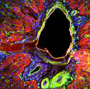
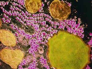
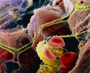
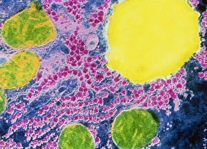
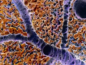
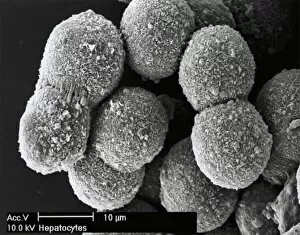
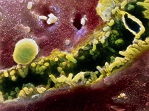
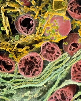
A hepatocyte, also known as a liver cell, is a vital component of the liver tissue. These specialized cells play a crucial role in maintaining the overall health and functionality of this essential organ. Under the powerful lens of a transmission electron microscope (TEM), one can observe the intricate laminae formed by hepatocytes. These layers create an organized structure within the liver tissue, allowing for efficient functioning. Unfortunately, certain conditions like cirrhosis can affect these remarkable cells. In light micrographs depicting cirrhosis of the liver, one can witness the detrimental impact on hepatocytes. The images reveal distorted and damaged cells that are unable to perform their duties effectively. In TEM images showcasing liver tissue affected by cirrhosis, further details emerge about how this condition disrupts normal cellular architecture. The once-organized arrangement becomes disrupted and irregular due to fibrosis and scarring caused by chronic inflammation. The importance of understanding hepatocytes' structure extends to studying liver portal triads - areas where hepatic arteries, bile ducts, and hepatic veins converge. Light micrographs provide insights into these complex structures within healthy liver tissue. To gain even more insight into hepatocyte morphology at a microscopic level, false-color TEM images offer enhanced visualization. These vibrant representations allow researchers to study various components within these cells with greater clarity. Additionally, scanning electron microscopy (SEM) provides colored views of both hepatocytes and bile canaliculi - small channels through which bile flows within the liver. This technique helps researchers examine not only individual cells but also their interconnections in detail. By combining different imaging techniques such as TEM and SEM with traditional light microscopy approaches, scientists continue to unravel mysteries surrounding hepatocytes' functions and dysfunctions alike. Understanding these fundamental units is crucial for developing treatments targeting diseases affecting this vital organ system.