Fraud Blocker Skip to main content

Gastrointestinal Collection

"Gastrointestinal

Background imageGastrointestinal Collection: Deer anatomy, artwork

Deer anatomy, artwork
Deer anatomy. Computer artwork showing the internal anatomy of a deer (family Cervidae). Anatomical features shown here include the skeleton, lungs, intestines, bladder

Background imageGastrointestinal Collection: Giardia lamblia protozoa, micrograph

Giardia lamblia protozoa, micrograph
Giardia lamblia protozoa, light micrograph. These single-celled parasitic protozoa infect the intestinal tract and are most common in tropical regions

Background imageGastrointestinal Collection: Intestinal villi, SEM

Intestinal villi, SEM
Intestinal villi. Coloured scanning electron micrograph (SEM) of a section through a part of the small intestine, showing the villi that cover its inner surface

Background imageGastrointestinal Collection: Gastric antral vascular ectasia C016 / 8328

Gastric antral vascular ectasia C016 / 8328
Gastric antral vascular ectasia. Endoscopic view of the antrum (final portion of stomach), affected by gastric antral vascular ectasia (GAVE)

Background imageGastrointestinal Collection: Colon cancer, endoscopic view C016 / 8308

Colon cancer, endoscopic view C016 / 8308
Colon cancer. Endoscopic view of a malignant (cancerous) adenocarcinoma (upper right) in a patients ascending colon (large intestine)

Background imageGastrointestinal Collection: Severe gastritis, endoscope view C016 / 3898

Severe gastritis, endoscope view C016 / 3898
Severe gastritis, endoscope view. Gastritis is inflammation of the stomach lining. It causes pain on eating or drinking, and may cause nausea and vomiting

Background imageGastrointestinal Collection: Goblet cells

Goblet cells. Coloured transmission electron micrograph (TEM) of a section through goblet cells in the lining of the small intestine, part of the digestive tract. They are full of mucus (yellow)

Background imageGastrointestinal Collection: Cow anatomy, artwork

Cow anatomy, artwork
Cow anatomy. Computer artwork showing the internal anatomy of a domesticated cow (Bos primigenius). Anatomical features shown here include the skeleton, lungs, intestines, bladder

Background imageGastrointestinal Collection: HK97 bacteriophage capsid

HK97 bacteriophage capsid, molecular model. Bacteriophages are viruses that infect bacteria, in this case enterobacteria such as E. coli (Escherichia coli), with the phage head shown here

Background imageGastrointestinal Collection: HK97 bacteriophage procapsid

HK97 bacteriophage procapsid. Molecular model showing the structure of the prohead-I procapsid of the HK97 bacteriophage. Bacteriophages are viruses that infect bacteria

Background imageGastrointestinal Collection: Intestinal veins, 1825 artwork

Intestinal veins, 1825 artwork
Intestinal veins. Dissection of the abdominal cavity showing the inferior mesenteric vein (blue) that drains blood from the large intestine

Background imageGastrointestinal Collection: Liver and stomach nerves, 1825 artwork

Liver and stomach nerves, 1825 artwork
Liver and stomach nerves. Extensive network of nerves (white) supplying the liver (upper left) and stomach (lower right). These nerves networks (plexuses)

Background imageGastrointestinal Collection: Intestinal arteries, 1825 artwork

Intestinal arteries, 1825 artwork
Intestinal arteries. Dissection of the abdomen, showing the inferior mesenteric artery (red) and its branches that supply oxygenated blood to the large intestine

Background imageGastrointestinal Collection: Cytoplasmic polyhedrosis virus capsid

Cytoplasmic polyhedrosis virus capsid, molecular model. Part of the Cypovirus genus and invariably fatal, this insect virus is transmitted by contamination of leaves eaten (examples include silkworms)

Background imageGastrointestinal Collection: Healthy spleen, artwork

Healthy spleen, artwork
Healthy spleen, computer artwork

Background imageGastrointestinal Collection: Colitis, light micrograph

Colitis, light micrograph
Colitis. Light micrograph of a section through colon (large intestine) tissue affected by bacterial infection, causing injury to the mucosal lining, which has resulted in inflammation (colitis)

Background imageGastrointestinal Collection: Ischaemic bowel, light micrograph

Ischaemic bowel, light micrograph
Ischaemic bowel. Light micrograph of a section through a portion of intestine that has been damaged by an interruption of the blood supply, or ischaemia

Background imageGastrointestinal Collection: Reovirus particles, TEM C016 / 9443

Reovirus particles, TEM C016 / 9443
Reovirus particles. Coloured transmission electron micrograph (TEM) of numerous reovirus particles (virions, round). Reoviruses affect the gastrointestinal system and respiratory tract

Background imageGastrointestinal Collection: Reovirus particles, TEM C016 / 9444

Reovirus particles, TEM C016 / 9444
Reovirus particles. Coloured transmission electron micrograph (TEM) of numerous reovirus particles (virions, blue). Reoviruses affect the gastrointestinal system and respiratory tract

Background imageGastrointestinal Collection: Total mesorectal excision, artwork C016 / 6996

Total mesorectal excision, artwork C016 / 6996
Total mesorectal excision (TME). Artwork of the pelvis and the area of the colon and rectum that is removed in total mesorectal excision

Background imageGastrointestinal Collection: Giardia protozoan, SEM C016 / 9398

Giardia protozoan, SEM C016 / 9398
Giardia protozoan. Coloured scanning electron micrograph (SEM) of a Giardia sp. trophozoite protozoan (yellow) attached to the lining of a human intestine

Background imageGastrointestinal Collection: Intestinal brush border, TEM

Intestinal brush border, TEM
Intestinal brush border. Transmission electron micrograph (TEM) of intestinal absorptive cells sectioned horizontally at their apex to show the surface microvilli (round)

Background imageGastrointestinal Collection: Gastritis, light micrograph C016 / 0508

Gastritis, light micrograph C016 / 0508
Gastritis. Light micrograph of a section through an inflamed human stomach (gastritis), showing an abnormal accumulation of white blood cells (leucocytes)

Background imageGastrointestinal Collection: Appendix, light micrograph C016 / 0510

Appendix, light micrograph C016 / 0510
Appendix. Light micrograph of a section through a human appendix. The appendix is a blind-ending small diameter tubular extension of the gut, arising from the caecum of the large bowel

Background imageGastrointestinal Collection: Gastritis, light micrograph C016 / 0507

Gastritis, light micrograph C016 / 0507
Gastritis. Light micrograph of a section through an inflamed human stomach (gastritis), showing an abnormal accumulation of white blood cells (leucocytes)

Background imageGastrointestinal Collection: Colon polyp, light micrograph C016 / 0512

Colon polyp, light micrograph C016 / 0512
Colon polyp. Light micrograph of a section through part of a tubulovillous adenoma of the mucosa of the large bowel (colon)

Background imageGastrointestinal Collection: Colon, light micrograph C016 / 0516

Colon, light micrograph C016 / 0516
Colon. Light micrograph of a section through the mucosa (inner epithelial lining) of the colon (large bowel), showing the typical arrangement of invaginations of columnar epithelial cells forming

Background imageGastrointestinal Collection: Islet of Langerhans, light micrograph C016 / 0514

Islet of Langerhans, light micrograph C016 / 0514
Islet of Langerhans. Light micrograph of a section through an islet of Langerhans in a pancreas. Scattered throughout the pancreas, the islets are endocrine cells that secrete a range of hormones

Background imageGastrointestinal Collection: Bowel cancer, light micrograph C016 / 0511

Bowel cancer, light micrograph C016 / 0511
Bowel cancer. Light micrograph of a section through a carcinoid tumour in the small bowel. The tumour has invaded the submucosa of the bowel

Background imageGastrointestinal Collection: Islet of Langerhans, light micrograph C016 / 0513

Islet of Langerhans, light micrograph C016 / 0513
Islet of Langerhans. Light micrograph of a section through an islet of Langerhans in a pancreas. Scattered throughout the pancreas, the islets are endocrine cells that secrete a range of hormones

Background imageGastrointestinal Collection: Enema and stomach pump, circa 1870 C018 / 0360

Enema and stomach pump, circa 1870 C018 / 0360
Nineteenth century enema and stomach pump, made from wood, metal, and rubber. An enema is a syringe-like device used to evacuate the lower intestinal tract through introducing fluids to the rectum

Background imageGastrointestinal Collection: Enema and stomach pump, circa 1880 C018 / 0361

Enema and stomach pump, circa 1880 C018 / 0361
Nineteenth century enema and stomach pump in a wooden case made from brass, wood, and ivory. An enema is a syringe-like device used to evacuate the lower intestinal tract through introducing fluids

Background imageGastrointestinal Collection: Weiss patent syringe, circa 1850 C018 / 0362

Weiss patent syringe, circa 1850 C018 / 0362
Weiss patent syringe for use as enema or stomach pump. The components of this apparatus are made from brass and ivory. It dates from around 1850, England

Background imageGastrointestinal Collection: Enema and stomach pump, circa 1870 C018 / 0374

Enema and stomach pump, circa 1870 C018 / 0374
Nineteenth century enema and stomach pump made from wood, metal, and rubber (in a wooden case). An enema is a syringe-like device used to evacuate the lower intestinal tract through introducing

Background imageGastrointestinal Collection: Norwalk virus capsid, molecular model

Norwalk virus capsid, molecular model. This norovirus, which causes a viral form of gastroenteritis, is transmitted from person-to-person or through contaminated food

Background imageGastrointestinal Collection: Simian rotavirus capsid, molecular model

Simian rotavirus capsid, molecular model. This virus is named for its ability to infect the higher primates (simians). Rotaviruses, transmitted by faecal-oral contact


All Professionally Made to Order for Quick Shipping

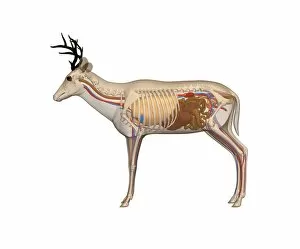
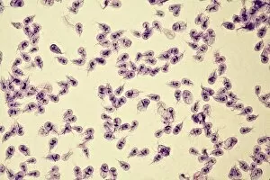
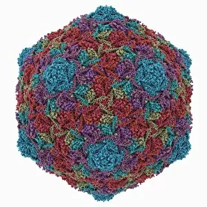
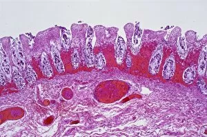
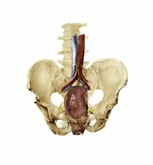
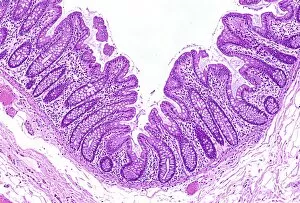
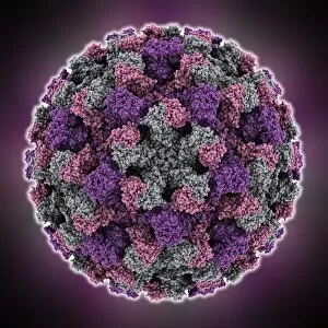
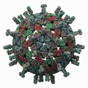
"Gastrointestinal: Exploring the Intricacies of Digestion Through Art and Anatomy" Delve into the fascinating world anatomy through a captivating blend of artwork and scientific exploration. From dogs to deer, cows to humans, this diverse collection showcases the complexity and beauty of our digestive systems. Intricate Dog Anatomy: Uncover the intricate network of organs that make up a dog's gastrointestinal system. Discover how each component plays a vital role in digestion, ensuring optimal nutrient absorption for our furry friends. Artistic Deer Anatomy: Immerse yourself in stunning artwork depicting the inner workings of a deer's digestive system. Marvel at nature's design as you witness how these majestic creatures process their food with precision. Intestinal Villi Under SEM: Journey deep into microscopic territory as scanning electron microscopy reveals the mesmerizing structure of intestinal villi. These tiny finger-like projections increase surface area, enabling efficient nutrient absorption within our own bodies. Gastric Antral Vascular Ectasia C016/8328: Explore an intriguing medical condition known as gastric antral vascular ectasia (GAVE). Delve into its visual representation and learn about its impact on blood vessels within the stomach lining. Giardia Lamblia Protozoa Micrograph: Peer through a microscope lens to observe Giardia lamblia protozoa – parasites responsible for causing giardiasis. Witness their unique appearance and understand their detrimental effects on human gastrointestinal health. Cow Anatomy Transformed Into Artwork: Admire stunning artistic renditions showcasing cow anatomy from an entirely new perspective. Gain insight into how these gentle giants process plant-based diets efficiently while maintaining overall health. Artistic Intestinal Villi Depiction: Step back from reality and immerse yourself in imaginative artwork capturing the essence of intestinal villi. Let your imagination run wild as you envision these crucial structures working tirelessly behind-the-scenes to support digestion. Male Anatomy Reimagined: Witness the male gastrointestinal system transformed into captivating artwork.