Fraud Blocker Skip to main content

Fruiting Body Collection (page 3)

"Fruiting Body: A Fascinating World of Mushroom and Slime Moulds" Nature's artistic creations never cease to amaze us

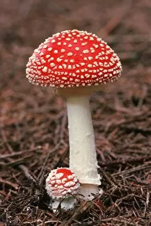
Background imageFruiting Body Collection: Fly agaric (Amanita muscaria) fungus C014 / 3019

Fly agaric (Amanita muscaria) fungus C014 / 3019
Fly agaric (Amanita muscaria) fungus. These poisonous mushrooms are the fruiting bodies of the Amanita muscaria fungus. The caps have long been dried

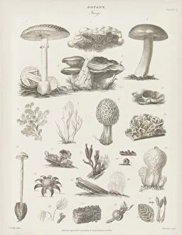
Background imageFruiting Body Collection: Fungal fruiting bodies, 19th century C015 / 6087

Fungal fruiting bodies, 19th century C015 / 6087
Fungal fruiting bodies, 19th-century artwork. The shape of fungal fruiting bodies can range from capped and gilled toadstools and mushrooms (top), to bracket fungi (upper centre), morels (centre)

Background imageFruiting Body Collection: Mycena epipterygia mushroom C018 / 2535

Mycena epipterygia mushroom C018 / 2535
Mycena epipterygia mushroom

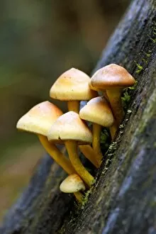
Background imageFruiting Body Collection: Sulphur tuft (Hypholoma fasciculare) C018 / 2565

Sulphur tuft (Hypholoma fasciculare) C018 / 2565
Sulphur tuft (Hypholoma fasciculare) mushrooms

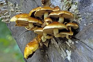
Background imageFruiting Body Collection: Sulphur tuft (Hypholoma fasciculare) C018 / 2534

Sulphur tuft (Hypholoma fasciculare) C018 / 2534
Sulphur tuft (Hypholoma fasciculare) mushrooms

Background imageFruiting Body Collection: Scleroderma citrinum mushroom C018 / 2538

Scleroderma citrinum mushroom C018 / 2538
Common earthball (Scleroderma citrinum) mushroom

Background imageFruiting Body Collection: Ash dieback disease fungus, SEM C017 / 8028

Ash dieback disease fungus, SEM C017 / 8028
Ash dieback disease fungus. Coloured scanning electron micrograph (SEM) of the surface of a fruiting body of the fungus Chalara fraxinea

Background imageFruiting Body Collection: Ash dieback disease fungus, SEM C017 / 8026

Ash dieback disease fungus, SEM C017 / 8026
Ash dieback disease fungus. Coloured scanning electron micrograph (SEM) of a fruiting body of the fungus Chalara fraxinea, also known as Hymenoscyphus pseudoalbidus in its fruiting stage

Background imageFruiting Body Collection: Ash dieback disease fungus, SEM C017 / 8027

Ash dieback disease fungus, SEM C017 / 8027
Ash dieback disease fungus. Coloured scanning electron micrograph (SEM) of fruiting bodies of the fungus Chalara fraxinea, also known as Hymenoscyphus pseudoalbidus in its fruiting stage

Background imageFruiting Body Collection: Shaggy pholiota (Pholiota aurivella) C018 / 2566

Shaggy pholiota (Pholiota aurivella) C018 / 2566
Shaggy pholiota (Pholiota aurivella) mushrooms

Background imageFruiting Body Collection: Beechwood sickener (Russula nobilis) C018 / 2532

Beechwood sickener (Russula nobilis) C018 / 2532
Beechwood sickener (Russula nobilis) mushroom

Background imageFruiting Body Collection: Shaggy pholiota (Pholiota aurivella) C018 / 2533

Shaggy pholiota (Pholiota aurivella) C018 / 2533
Shaggy pholiota (Pholiota aurivella) mushrooms

Background imageFruiting Body Collection: Hypholoma mushrooms C018 / 2568

Hypholoma mushrooms C018 / 2568
Hypholoma mushrooms. Sulphur tuft (Hypholoma fasciculare, left) and brick cap (Hypholoma sublateritium, right) mushrooms growing on a tree stump

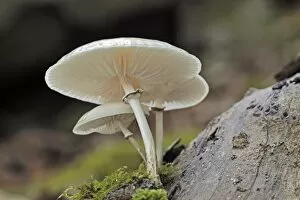
Background imageFruiting Body Collection: Porcelain fungus (Oudemansiella mucida) C018 / 2564

Porcelain fungus (Oudemansiella mucida) C018 / 2564
Porcelain fungus (Oudemansiella mucida) mushroom

Background imageFruiting Body Collection: Mycena epipterygia mushroom C018 / 2536

Mycena epipterygia mushroom C018 / 2536
Mycena epipterygia mushroom

Background imageFruiting Body Collection: Scutiger pes-caprae-sabatera mushrooms C016 / 3446

Scutiger pes-caprae-sabatera mushrooms C016 / 3446
Scutiger pes-caprae-sabatera mushrooms, artwork

Background imageFruiting Body Collection: Caesars mushrooms (Amanita caesarea)

Caesars mushrooms (Amanita caesarea), artwork

Background imageFruiting Body Collection: Puffball (Lycoperdon perlatum) mushrooms C016 / 3434

Puffball (Lycoperdon perlatum) mushrooms C016 / 3434
Puffball (Lycoperdon perlatum) mushrooms, artwork

Background imageFruiting Body Collection: Fairy ring (Marasmius oreades) mushrooms C016 / 3433

Fairy ring (Marasmius oreades) mushrooms C016 / 3433
Fairy ring (Marasmius oreades) mushrooms, artwork

Background imageFruiting Body Collection: Lactarius deliciosus mushrooms, artwork C016 / 3367

Lactarius deliciosus mushrooms, artwork C016 / 3367
Saffron milk cap (Lactarius deliciosus) mushrooms, artwork

Background imageFruiting Body Collection: Hygrophorus russula mushrooms, artwork C016 / 3364

Hygrophorus russula mushrooms, artwork C016 / 3364
Hygrophorus russula mushrooms, artwork

Background imageFruiting Body Collection: Aspergillus fungus, artwork C013 / 4613

Aspergillus fungus, artwork C013 / 4613
Aspergillus fungus. Computer artwork of the fruiting body (conidiophore) of a typical mould (Aspergillus sp.), showing spores (conidia, round)

Background imageFruiting Body Collection: Weeping widow mushrooms C013 / 5107

Weeping widow mushrooms C013 / 5107
Weeping widow mushrooms (Lacrymaria velutina). Photographed in autumn, in Normandy, France

Background imageFruiting Body Collection: Giant puffball fungus in a basket C013 / 5108

Giant puffball fungus in a basket C013 / 5108
Giant puffball fungus (Calvatia gigantea) in a basket with a shallot and a bulb of garlic. Photographed in autumn, in Cornwall, UK

Background imageFruiting Body Collection: Common ink cap mushrooms C013 / 5106

Common ink cap mushrooms C013 / 5106
Common ink cap mushrooms (Coprinopsis atramentaria). Photographed in autumn, in Normandy, France

Background imageFruiting Body Collection: Russula emetica fungus

Russula emetica fungus growing on moss. Known as the sickener mushroom, this poisonous fungus is usually found growing in woodland areas on the forest floor. Photographed in the UK

Background imageFruiting Body Collection: Rust fungus, SEM

Rust fungus, SEM
Rust fungus. Coloured scanning electron micrograph (SEM) of spores of the mint rust fungus (Puccinia menthae, orange) on the surface of a peppermint leaf (Mentha piperita)

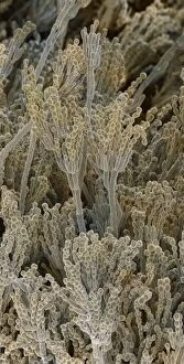
Background imageFruiting Body Collection: Penicillium fungal spores, SEM

Penicillium fungal spores, SEM
Penicillium fungal spores. Coloured scanning electron micrograph (SEM) of fruiting bodies of the fungus Penicillium roqueforti

Background imageFruiting Body Collection: Bonnet

Bonnet
Collected during the archaelogical excavation at Christ Church, Spitalfields, London, 1984-1986

Background imageFruiting Body Collection: Various fungi

Various fungi
Plate 25 from Le Regne Vegetal, Vol 12, Hort. Atlas 1870. Held in the Botany Library at the Natural History Museum, London. Illustration entitled Cryptogames alimentaires

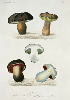
Background imageFruiting Body Collection: Bolete sp. bolete mushrooms

Bolete sp. bolete mushrooms
Plate 24 from Le Regne Vegetal plantes agricoles, Vol 8, depicting various types and views of bolete mushrooms

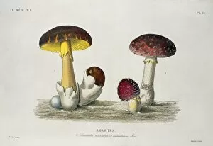
Background imageFruiting Body Collection: Amanita sp. amanita mushrooms

Amanita sp. amanita mushrooms
Plate 10 depicting two different species of amanite mushrooms from Le Regne Vegetal plantes agricoles, Vol 8

Background imageFruiting Body Collection: Balanus tintinnabulum, balanidae barnacles

Balanus tintinnabulum, balanidae barnacles
Plate 1 by George Sowerby from Charles Darwins Monograph on the sub-class Cirripedia, with figures of all the species: the Balan

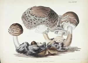
Background imageFruiting Body Collection: Agaricus rachodes

Agaricus rachodes. Plate XXXVIII taken from Illustrations of British Mycology by Hussey, Anna Maria

Background imageFruiting Body Collection: Clathrus ruber, latticed stinkhorn

Clathrus ruber, latticed stinkhorn
Illustration from Nova Plantarum (1729) by Pietro Antonio Micheli, of Type Specimens of plants named by Carl Linnaeus (1707-1778)

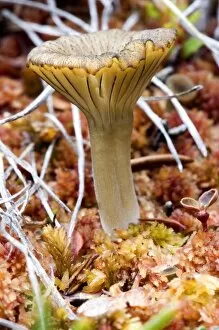
Background imageFruiting Body Collection: An arctic mushroom (approximately 3cm tall) emerges from wet tundra - Arctic National

An arctic mushroom (approximately 3cm tall) emerges from wet tundra - Arctic National Wildlife Refuge, Alaska

Background imageFruiting Body Collection: Parasol Fungus (Macrolepiota procera) in Grassland. England, UK

Parasol Fungus (Macrolepiota procera) in Grassland. England, UK

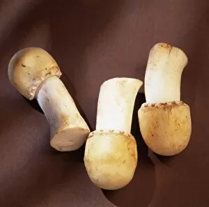
Background imageFruiting Body Collection: Horse mushrooms

Horse mushrooms (Agaricus arvensis). This fungus is an excellent food source, having a mushroomy taste and an aniseed smell. The white caps yellow slightly on ageing or when bruised

Background imageFruiting Body Collection: Shiitake mushroom

Shiitake mushroom (Lentinula edodes). Also known as black forest mushrooms, this fungus has been cultivated in the eastern Asia for over 650 years

Background imageFruiting Body Collection: Earth ball fungus, light micrograph

Earth ball fungus, light micrograph
Earth ball fungus. Light micrograph of a section through tissue from the fruiting body of an earth ball (Scleroderma vulgare) mushroom

Background imageFruiting Body Collection: Boletus fungus, light micrograph

Boletus fungus, light micrograph
Boletus fungus. Light micrograph of a section through tissue from the fruiting body of a Boletus sp. mushroom, showing the hymenium (dark red) of the fruiting body

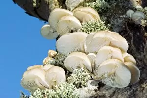
Background imageFruiting Body Collection: Porcelain fungus

Porcelain fungus (Oudemansiella mucida). Fruiting bodies of the porcelain fungus growing on the trunk of a dead beech tree (Fagus sylvaticus)

Background imageFruiting Body Collection: Gill fungus (Lyophyllum decastes)

Gill fungus (Lyophyllum decastes) occurs in dense clusters. The word decastes means a company of ten men. The gills are covered in basidia bearing spores

Background imageFruiting Body Collection: Beefsteak Fungus (Fistulina hepatica)

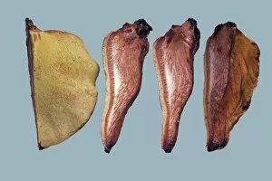
Beefsteak Fungus (Fistulina hepatica). This bracket fungus belonging to the homobasidiomycetes grows on the bark of living oak and sweet chestnut trees

Background imageFruiting Body Collection: Magic mushrooms (Psilocybe semilanceata)

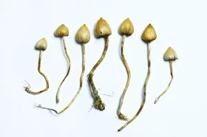
Magic mushrooms (Psilocybe semilanceata). These fungi, commonly known as liberty caps, contain the psychoactive compound psilocybin, which produces a hallucinogenic effect when ingested

Background imageFruiting Body Collection: Tremella mesenterica

Tremella mesenterica
Yellow brain fungus (Tremella mesenterica) growing on a dead gorse bush. The fruiting body is soft, gelatinous and translucent with brain like lobes

Background imageFruiting Body Collection: Common Earthball (Scleroderma citrinum)

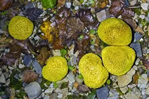
Common Earthball (Scleroderma citrinum). The fruiting body is hemispherical with a tough outer layer covered in scales. Inside the ball is a purple-brown mass (gleba)

Background imageFruiting Body Collection: Ladybird on fungus

Ladybird on fungus. Seven-spot ladybird (Coccinella septem-punctata) climbing up the stalk of a fungal fruiting body. This is a wood-rotting fungus growing on a dead tree stump


All Professionally Made to Order for Quick Shipping

"Fruiting Body: A Fascinating World of Mushroom and Slime Moulds" Nature's artistic creations never cease to amaze us, especially when it comes to the diverse forms and structures found in fruiting bodies. From the iconic Fly agaric mushrooms with their vibrant red caps speckled with white dots, to the intricate patterns of mushroom gills observed under a scanning electron microscope (SEM), these organisms captivate our imagination. But it doesn't stop there. The world of fruiting bodies extends beyond mushrooms alone. Take, for instance, the fruiting bodies of Rhizopus oligosporus, a type of mold used in food fermentation processes like tempeh production. These tiny structures play a crucial role in transforming ingredients into delicious culinary delights. Moving on from molds to lichens, we encounter the enchanting Cup lichen (Cladonia floerkeana). Its cup-shaped fruiting body adds an ethereal touch to forest floors or rocky landscapes where it thrives. And then there are slime moulds – peculiar organisms that blur the line between fungi and protists. In Buckinghamshire, England's woodlands during November, Stemonitopsis typhina sporangia can be spotted growing on bark edges. These slimy masses release spores as they mature—a mesmerizing sight captured through focus stacking photography techniques. Another slime mould species found in Buckinghamshire is Metatrichia floriformis. Its line of split-open sporangia reveals delicate spore-filled interiors against a backdrop of winter scenery—nature's own miniature explosions frozen in time. Zooming even closer into this microscopic world brings us Lamproderma scintillans—a slime mould boasting 1mm tall sporangia that shimmer like stars under magnification—an awe-inspiring spectacle hidden within nature's nooks and crannies.