Home > Science > SEM
Rust fungus, SEM

![]()
Wall Art and Photo Gifts from Science Photo Library
Rust fungus, SEM
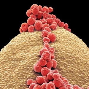
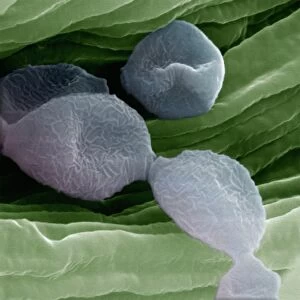
Rust fungus. Coloured scanning electron micrograph (SEM) of spores of the mint rust fungus (Puccinia menthae, orange) on the surface of a peppermint leaf (Mentha piperita). The reproductive spores have been released from a fruiting body (pink), which ruptures the epidermal surface of the leaf in the later stages of fungal infection. The mint rust can cause severe yield losses in commercial mint crops. Magnification: x130 at 6x7cm size
Science Photo Library features Science and Medical images including photos and illustrations
Media ID 9194093
© POWER AND SYRED/SCIENCE PHOTO LIBRARY
Bodies Diseased Diseases Fruiting Body Fungi Infected Infection Infectious Lesion Mold Mould Mouldy Mycology Parasite Parasitic Pathogens Pathology Reproducing Reproduction Reproductive Spore Spores Structures Mentha Piperita Peppermint
EDITORS COMMENTS
This print showcases the intricate world of rust fungus, captured through a coloured scanning electron micrograph (SEM). The image reveals the spores of the mint rust fungus, Puccinia menthae, in vibrant orange hues. These reproductive spores have been released from a fruiting body that ruptures the surface of a peppermint leaf (Mentha piperita) during later stages of fungal infection. The mint rust fungus is notorious for causing significant yield losses in commercial mint crops. Its parasitic nature and infectious properties make it a formidable pathogen to battle against. This SEM image provides an up-close look at the structures and bodies involved in this destructive process. The magnification level of x130 allows us to appreciate the minute details present on this 6x7cm-sized print. From lesions and moldy patches to reproductive spores and fruiting bodies, every element tells its own story within this microscopic world. As we delve into this mesmerizing realm of mycology and pathology, we gain insight into how diseases can impact plant life on such a minuscule scale. This photograph serves as both an educational tool for understanding these complex interactions between fungi and plants, as well as a visually stunning piece that highlights the beauty found even in disease-ridden environments.
MADE IN THE UK
Safe Shipping with 30 Day Money Back Guarantee
FREE PERSONALISATION*
We are proud to offer a range of customisation features including Personalised Captions, Color Filters and Picture Zoom Tools
SECURE PAYMENTS
We happily accept a wide range of payment options so you can pay for the things you need in the way that is most convenient for you
* Options may vary by product and licensing agreement. Zoomed Pictures can be adjusted in the Basket.