
Flagellum Collection
"Exploring the Fascinating World of Flagellum: From Cell Types to Artwork" Flagellum, a remarkable feature found in various cell types


All Professionally Made to Order for Quick Shipping
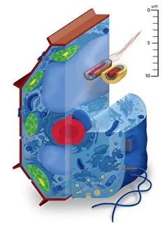
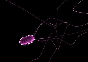
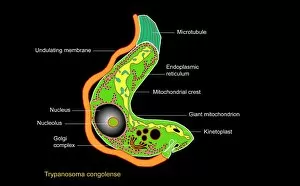
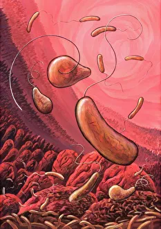
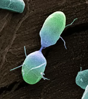
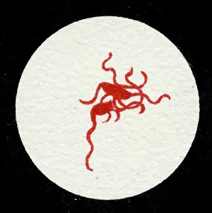
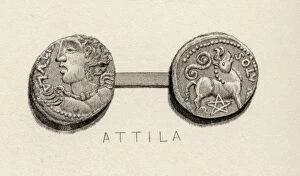
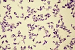
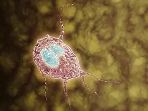
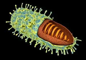
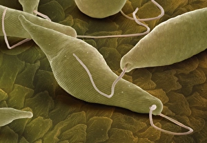
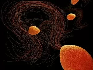
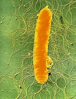
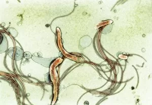
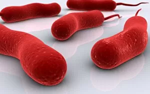
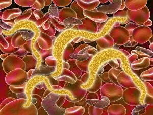
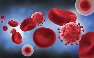
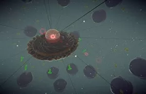
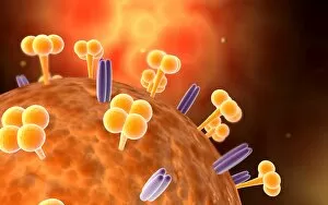
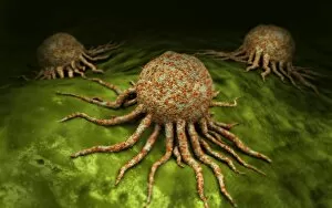
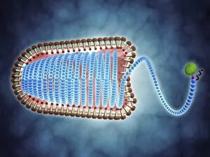
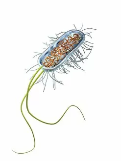
"Exploring the Fascinating World of Flagellum: From Cell Types to Artwork" Flagellum, a remarkable feature found in various cell types, has captivated scientists and artists alike. In the microscopic realm, flagellate bacteria such as the E. Coli bacterium showcase their intricate propulsion system through these whip-like appendages. The Trypanosome protozoan, depicted in stunning artwork, mesmerizes with its elegant flagella that enable it to navigate through diverse environments. Artistic renditions also bring attention to the role of flagella in disease-causing organisms like Cholera bacteria. Through vivid illustrations, we witness how these pathogens employ their flagella for mobility and colonization within our bodies. Similarly, electron microscopy unveils captivating images of Salmonella bacterium dividing and Salmonella typhimurium bacteria with their distinct flagellar structures. Intriguingly colored transmission electron micrographs reveal Escherichia coli bacteria adorned with vibrant flagella—a sight that showcases both scientific precision and artistic beauty. Delving into history, lithographs from 1906 depict a colony of Salmonella Typhi exhibiting Bacilli with prominent flagella—an early exploration into understanding bacterial morphology. Beyond scientific realms, ancient engravings portray the symbolic significance of the "Scourge, " an alternative name for the flagellum. These depictions highlight how humans have long been fascinated by this unique cellular structure's potential impact on life itself. Just as there are countless varieties of eels inhabiting our oceans' depths—each possessing different adaptations—the world presents us with an equally diverse array of forms and functions across species. Whether observed under a microscope or immortalized through artistry throughout history, exploring the intricacies of this cellular appendage continues to inspire awe and curiosity among scientists and artists alike. So let us delve deeper into this captivating world where science meets art—a realm where tiny cells propel themselves forward, guided by the graceful movements of their flagella.