Fraud Blocker Skip to main content

Dissection Collection (page 7)

"Unveiling the Wonders Within

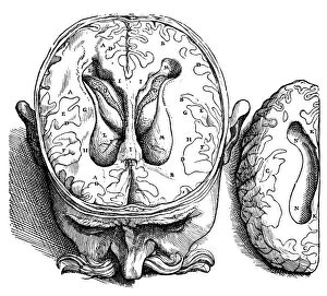
Background imageDissection Collection: Dissection of the brain (fig. 8). Woodcut from the seventh book of Andreas Vesalius De Humani

Dissection of the brain (fig. 8). Woodcut from the seventh book of Andreas Vesalius De Humani Corporis Fabrica
VESALIUS: BRAIN, 1543. Dissection of the brain (fig. 8). Woodcut from the seventh book of Andreas Vesalius De Humani Corporis Fabrica, published in 1543 at Basel, Switzerland

Background imageDissection Collection: Dissection of the brain (fig. 5). Woodcut from the seventh book of Andreas Vesalius De Humani

Dissection of the brain (fig. 5). Woodcut from the seventh book of Andreas Vesalius De Humani Corporis Fabrica
VESALIUS: BRAIN, 1543. Dissection of the brain (fig.5). Woodcut from the seventh book of Andreas Vesalius De Humani Corporis Fabrica, published in 1543 at Basel, Switzerland

Background imageDissection Collection: Dissection of the brain (fig. 6). Woodcut from the seventh book of Andreas Vesalius De Humani

Dissection of the brain (fig. 6). Woodcut from the seventh book of Andreas Vesalius De Humani Corporis Fabrica
VESALIUS: BRAIN, 1543. Dissection of the brain (fig.6). Woodcut from the seventh book of Andreas Vesalius De Humani Corporis Fabrica, published in 1543 at Basel, Switzerland

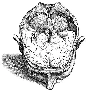
Background imageDissection Collection: Dissection of the brain (fig. 9). Woodcut from the seventh book of Andreas Vesalius De Humani

Dissection of the brain (fig. 9). Woodcut from the seventh book of Andreas Vesalius De Humani Corporis Fabrica, Basel
VESALIUS: BRAIN, 1543. Dissection of the brain (fig.9). Woodcut from the seventh book of Andreas Vesalius De Humani Corporis Fabrica, Basel, Switzerland, 1543

Background imageDissection Collection: Woodcut bloodletting chart by Johannes Wechtlin, 1517

Woodcut bloodletting chart by Johannes Wechtlin, 1517
BLOODLETTING CHART, 1517. Woodcut bloodletting chart by Johannes Wechtlin, 1517

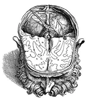
Background imageDissection Collection: Dissection of the brain (fig. 4). Woodcut from the seventh book of Andreas Vesalius De Humani

Dissection of the brain (fig. 4). Woodcut from the seventh book of Andreas Vesalius De Humani Corporis Fabrica
VESALIUS: BRAIN, 1543. Dissection of the brain (fig.4). Woodcut from the seventh book of Andreas Vesalius De Humani Corporis Fabrica, published in 1543 at Basel, Switzerland

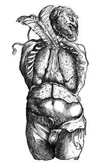
Background imageDissection Collection: Woodcut from the sixth book of Andreas Vesalius De Humani Corporis Fabrica

Woodcut from the sixth book of Andreas Vesalius De Humani Corporis Fabrica, published in 1543 at Basel, Switzerland
VESALIUS: THORACIC CAVITY. Woodcut from the sixth book of Andreas Vesalius De Humani Corporis Fabrica, published in 1543 at Basel, Switzerland

Background imageDissection Collection: Dissection of the brain (fig. 7). Woodcut from the seventh book of Andreas Vesalius De Humani

Dissection of the brain (fig. 7). Woodcut from the seventh book of Andreas Vesalius De Humani Corporis Fabrica
VESALIUS: BRAIN, 1543. Dissection of the brain (fig.7). Woodcut from the seventh book of Andreas Vesalius De Humani Corporis Fabrica published in 1543 at Basel, Switzerland

Background imageDissection Collection: Title page of Mondino de Luzzi (Mundinus) Anathomia, published by Martin Pollick

Title page of Mondino de Luzzi (Mundinus) Anathomia, published by Martin Pollick van Mellerstadt, Leipzig, 1493, showing anatomy demonstration

Background imageDissection Collection: Dissection in progress

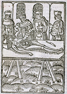
Dissection in progress
A dissection in progress. From " Le livre de la propriete des choses" (On the Properites of Things) written by the English Franciscan monk Bartholomew Glanville called Bartolomaeus Anglicus

Background imageDissection Collection: Title page of Andreas Vesalius De Humani Corporis Fabrica

Title page of Andreas Vesalius De Humani Corporis Fabrica, Basel, 1543, showing Vesalius dissecting a body

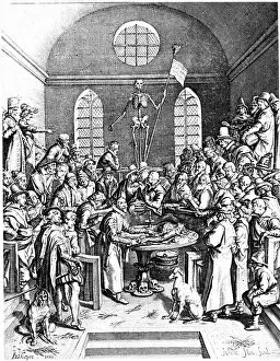
Background imageDissection Collection: Late 16th century anatomy theatre by Jacques de Gehyn the Elder

Late 16th century anatomy theatre by Jacques de Gehyn the Elder. From Dutch translation by Vopiscus Plemp of Barthelemy Cabrol " Anatomical Alphabet", Amsterdam, 1633. Engraving

Background imageDissection Collection: The second plate of the muscles

The second plate of the muscles. From Book II of Andreas Vesalius " De humani corporis fabrica", Basel, 1543. Engraving

Background imageDissection Collection: Twisted-pair cabling, side view

Twisted-pair cabling, side view

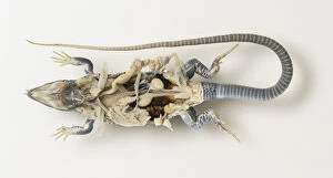
Background imageDissection Collection: Dissected lizard with intestines revealed, view from below

Dissected lizard with intestines revealed, view from below

Background imageDissection Collection: Dissected lab rat with intestines revealed, view from below

Dissected lab rat with intestines revealed, view from below

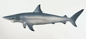
Background imageDissection Collection: Model of Spinner Shark (Carcharhinus brevipinna), side view

Model of Spinner Shark (Carcharhinus brevipinna), side view

Background imageDissection Collection: Human foot anatomy, artwork

Human foot anatomy, artwork
Human foot anatomy, computer artwork

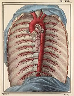
Background imageDissection Collection: Thoracic aorta, 1825 artwork

Thoracic aorta, 1825 artwork
Thoracic aorta. Dissection of the chest cavity with the heart and lungs and other organs removed, showing the thoracic portion of the aorta (red), the main body artery

Background imageDissection Collection: Intestinal veins, 1825 artwork

Intestinal veins, 1825 artwork
Intestinal veins. Dissection of the abdominal cavity showing the inferior mesenteric vein (blue) that drains blood from the large intestine

Background imageDissection Collection: Liver and stomach nerves, 1825 artwork

Liver and stomach nerves, 1825 artwork
Liver and stomach nerves. Extensive network of nerves (white) supplying the liver (upper left) and stomach (lower right). These nerves networks (plexuses)

Background imageDissection Collection: Intestinal arteries, 1825 artwork

Intestinal arteries, 1825 artwork
Intestinal arteries. Dissection of the abdomen, showing the inferior mesenteric artery (red) and its branches that supply oxygenated blood to the large intestine

Background imageDissection Collection: Chest lymph vessels, 1825 artwork

Chest lymph vessels, 1825 artwork
Chest lymph vessels. Dissection of the thoracic cavity showing lymphatic vessels (white) and nodes. The central chest lymph vessel is the thoracic canal, running parallel to the backbone (centre)

Background imageDissection Collection: Heart-lung anatomy, 1825 artwork

Heart-lung anatomy, 1825 artwork
Heart-lung anatomy. Axial section through the chest at the level of the fifth thoracic vertebra (T5). The front of the body is at top

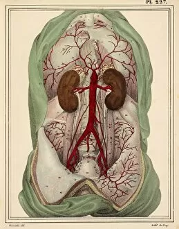
Background imageDissection Collection: Abdominal aorta, 1825 artwork

Abdominal aorta, 1825 artwork
Abdominal aorta. Dissection, with some organs removed, showing the abdominal portion of the aorta (red), the main body artery

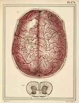
Background imageDissection Collection: Brain membrane vessels, 1825 artwork

Brain membrane vessels, 1825 artwork
Brain membrane vessels. Axial section through the skull showing the blood vessels (red) of two of the brain membranes, the arachnoid mater and pia mater

Background imageDissection Collection: Kidney anatomy, 1825 artwork

Kidney anatomy, 1825 artwork
Kidney anatomy. At top are the left and right kidneys (brown) with arteries (red), veins (blue), ureters (white) and adrenal glands (cream, on top of kidneys)

Background imageDissection Collection: Shark heart-gill anatomy, artwork C016 / 6847

Shark heart-gill anatomy, artwork C016 / 6847
Shark heart-gill anatomy. Artwork of the heart and gill anatomy of a shark, seen from the underside, with the head at right (teeth at centre right)

Background imageDissection Collection: Dissected mouse, illustration C018 / 0781

Dissected mouse, illustration C018 / 0781
Dissected mouse (Mus musculus), illustration

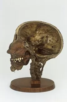
Background imageDissection Collection: Sectional dissected head, 19th century C017 / 0705

Sectional dissected head, 19th century C017 / 0705
Dissected cross section of a human head and neck with the brain removed. Anatomy of the skull, spine, teeth and sinuses can be seen. This artefact dates from the nineteenth century

Background imageDissection Collection: Female groin arteries, 1825 artwork

Female groin arteries, 1825 artwork
Female groin arteries. Dissections of a female groin to show areas supplied by the internal iliac artery (red) and its branches

Background imageDissection Collection: Lumbar spinal nerves, 1825 artwork

Lumbar spinal nerves, 1825 artwork
Lumbar spinal nerves. Lateral view of the lower vertebral column, showing nerves (white) of the sympathetic nervous system. The pelvic ilium bone has been removed

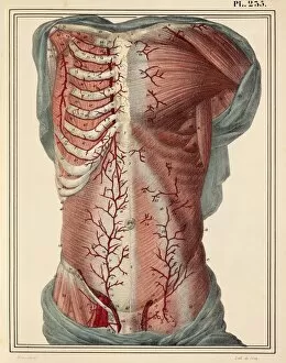
Background imageDissection Collection: Trunk arteries, 1825 artwork

Trunk arteries, 1825 artwork
Trunk arteries. Dissection showing the internal mammary arteries (red, in chest) and the epigastric arteries (red, in groin and abdomen), from a mans trunk (torso)

Background imageDissection Collection: Lower leg arteries, 1825 artwork

Lower leg arteries, 1825 artwork
Lower leg arteries. Anterior (frontal) view of the arteries (red) and muscles and ligaments of the lower leg and foot. At left are the superficial arteries

Background imageDissection Collection: Neck and shoulder arteries, 1825 artwork

Neck and shoulder arteries, 1825 artwork
Neck and shoulder arteries (red). Dissection showing the right carotid arteries of the neck, and the right subclavian artery of the shoulder

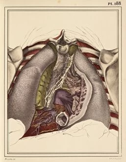
Background imageDissection Collection: Lung lymph vessels, 1825 artwork

Lung lymph vessels, 1825 artwork
Lung lymph vessels. Dissected right lung showing its lymph vessels (white). The dark bands are fissures between the different lobes of the lung

Background imageDissection Collection: Pulmonary nerve plexus, 1825 artwork

Pulmonary nerve plexus, 1825 artwork
Pulmonary nerve plexus. View from behind, with the thoracic spine removed, of the posterior pulmonary plexus, a confluence of nerves (white)

Background imageDissection Collection: Liver and stomach arteries, 1825 artwork

Liver and stomach arteries, 1825 artwork
Liver and stomach arteries. Dissection of the abdomen, showing the arteries (red) of the liver (brown, upper left), stomach (pink, upper centre), and the intestines (grey with yellow fat)

Background imageDissection Collection: Veins of the stomach, 1825 artwork

Veins of the stomach, 1825 artwork
Veins of the stomach. Abdominal dissection showing the veins (blue) of the stomach (centre) and the greater omentum (lower centre, yellow)

Background imageDissection Collection: Brain sinuses, 1825 artwork

Brain sinuses, 1825 artwork
Brain sinuses. Axial section through the head at the level of the base of the skull (front of head at top), showing the sinuses (blue) of the brains dura mater

Background imageDissection Collection: Intestinal lymph vessels, 1825 artwork

Intestinal lymph vessels, 1825 artwork
Intestinal lymph vessels. Dissection of the abdominal cavity showing lymphatic vessels (white) and nodes (lumps). The thoracic duct (top) connects with the intestinal lymph trunk (centre)

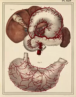
Background imageDissection Collection: Digestive organ arteries, 1825 artwork

Digestive organ arteries, 1825 artwork
Digestive organ arteries. Arteries (red) of various organs of the digestive system. At bottom are arteries of the posterior side of the stomach

Background imageDissection Collection: Femoral and foot arteries, 1825 artwork

Femoral and foot arteries, 1825 artwork
Femoral and foot arteries. Dissections showing the arteries (red) and muscles of the upper thigh and the foot. At centre right, near the bladder (white)

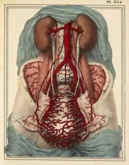
Background imageDissection Collection: Uterine arteries, 1825 artwork

Uterine arteries, 1825 artwork
Uterine arteries. Dissection showing the arteries (red) of the uterus (lower centre) of a woman who died six days after giving birth. The ovaries and fallopian tubes are either side

Background imageDissection Collection: Arteries of the thigh, 1825 artwork

Arteries of the thigh, 1825 artwork
Arteries of the thigh. Dissection showing the arteries (red) and muscles of the anterior (frontal) aspect of the thigh. The main artery in each leg is the femoral artery

Background imageDissection Collection: Internal body organs, 1825 artwork

Internal body organs, 1825 artwork
Internal body organs. Dissection showing the internal organs of the chest and abdomen. The overlying tissues have been removed to show (from top) the lungs

Background imageDissection Collection: Vena cavae veins, 1825 artwork

Vena cavae veins, 1825 artwork
Vena cavae veins. Dissection showing the veins (blue) associated with the vertebral column and heart. The superior (top) and inferior vena cava drain blood to the heart (centre, removed)

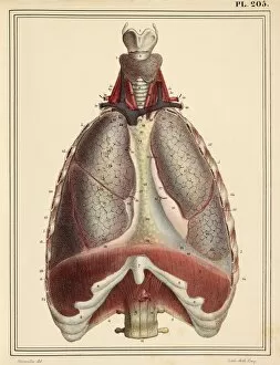
Background imageDissection Collection: Lungs and diaphragm, 1825 artwork

Lungs and diaphragm, 1825 artwork
Lungs and diaphragm. Anterior view of the chest, dissected vertically to reveal the lobes of the lungs, the diaphragm (brown, across bottom)


All Professionally Made to Order for Quick Shipping

"Unveiling the Wonders Within: Exploring the Intricacies of Dissection" Step into a world where art and science intertwine as we delve into the captivating realm of dissection. From Leonardo da Vinci's meticulous study of skull anatomy to Rembrandt's haunting depiction of anatomical dissections, these masterpieces offer us a glimpse into the intricate workings of the human body. Witness how Leonardo da Vinci's unrivaled genius comes alive in his Skull Anatomy, revealing every contour and detail with astonishing precision. Marvel at his ability to unravel the mysteries hidden beneath our skin, showcasing not only beauty but also scientific accuracy. Venture further and explore the beating heart, an organ that symbolizes life itself. Discover its complex network of blood vessels intricately depicted by skilled artists throughout history. These illustrations serve as a testament to our relentless pursuit in understanding this vital organ. Continue your journey through time and witness groundbreaking works like De humani corporis fabrica by Andreas Vesalius, which revolutionized anatomical studies during the Renaissance period. Delve deeper into Rembrandt's Dissection series, capturing both fascination and unease surrounding this practice. Feast your eyes on detailed drawings depicting musculature from various angles; each stroke meticulously crafted to showcase every fiber and sinew. These artworks remind us that beneath our skin lies a symphony of muscles working tirelessly together. Immerse yourself in The Anatomy Lesson of Dr Nicolaes Tulp by Rembrandt Harmensz van Rijn—a masterpiece that captures both scientific inquiry and artistic brilliance. Witness how physicians gather around their subject for an educational experience unlike any other. Finally, gaze upon breathtaking artwork illustrating arteries coursing through head, chest, male groin—each stroke telling stories untold until now. These visual narratives transport us back centuries ago when exploration was paramount in unlocking medical knowledge. Join us on this extraordinary journey where art meets science—a celebration of the human body's complexity and our relentless pursuit of knowledge.