Fraud Blocker Skip to main content

Digestive System Collection (page 4)

The digestive system is a complex network of organs and processes that allows our bodies to break down food and absorb nutrients

Background imageDigestive System Collection: Anatomy of liver, antero-visceral view

Anatomy of liver, antero-visceral view

Background imageDigestive System Collection: Conceptual image of human pancreas

Conceptual image of human pancreas

Background imageDigestive System Collection: Ureteropelvic junction (UPJ) obstruction in the kidney

Ureteropelvic junction (UPJ) obstruction in the kidney. UPJ is defined as an obstruction of the flow of urine from the renal pelvis to the proximal ureter

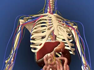
Background imageDigestive System Collection: Human skeleton showing digestive system and nervous system

Human skeleton showing digestive system and nervous system

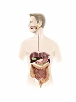
Background imageDigestive System Collection: Human digestive system

Human digestive system

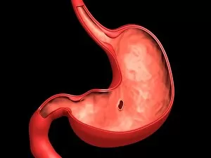
Background imageDigestive System Collection: Conceptual image of peptic ulcer in human stomach

Conceptual image of peptic ulcer in human stomach

Background imageDigestive System Collection: Conceptual image of human pancreas and stomach

Conceptual image of human pancreas and stomach

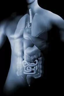
Background imageDigestive System Collection: Male body with internal organs

Male body with internal organs

Background imageDigestive System Collection: Human respiratory and digestive system

Human respiratory and digestive system
Medical illustration of human respiratory and digestive system

Background imageDigestive System Collection: 3D rendering of liver and stomach of human digestive system

3D rendering of liver and stomach of human digestive system

Background imageDigestive System Collection: Anatomy of a tracheoesophageal fistula

Anatomy of a tracheoesophageal fistula

Background imageDigestive System Collection: Medical illustration of the large intestine and small intestine

Medical illustration of the large intestine and small intestine, three dimensional view

Background imageDigestive System Collection: Human digestive system showing pancreas, duodenum, and gall bladder

Human digestive system showing pancreas, duodenum, and gall bladder
Human digestive system showing pancreas, duodenum, gall bladder, veins and arteries, isolated on white background

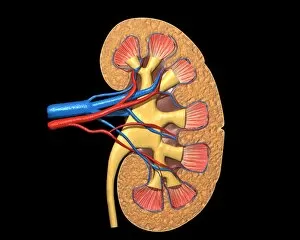
Background imageDigestive System Collection: Cross section of human kidney on black background

Cross section of human kidney on black background

Background imageDigestive System Collection: Anatomy of a childs full digestive system

Anatomy of a childs full digestive system, including mouth and nasal cross section. Other iinternal organs appear in half tone

Background imageDigestive System Collection: Male body standing, with full respiratory system superimposed

Male body standing, with full respiratory system superimposed
Human male body standing, with full respiratory system superimposed

Background imageDigestive System Collection: 3D rendering of human digestive system

3D rendering of human digestive system

Background imageDigestive System Collection: Anatomy of female body with organs

Anatomy of female body with organs

Background imageDigestive System Collection: Conceptual image of enterokinase

Conceptual image of enterokinase. Also known as enteropeptidase, it is an enzyme produced by cells of the duodenum and involved in human and animal digestion

Background imageDigestive System Collection: Conceptual image of human digestive system

Conceptual image of human digestive system showing gallbladder, pancrease, stomach and liver

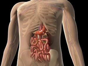
Background imageDigestive System Collection: Transparent view of human body showing kidney and intestines

Transparent view of human body showing kidney and intestines

Background imageDigestive System Collection: Hydronephrosis condition of the kidney

Hydronephrosis condition of the kidney
Hydronephrosis is a condition that typically occurs when one kidney becomes swollen due to the failure of normal drainage of urine from the kidney to the bladder

Background imageDigestive System Collection: Conceptual image of simple columnar epithelium

Conceptual image of simple columnar epithelium

Background imageDigestive System Collection: Anatomy of a childs full respiratory system

Anatomy of a childs full respiratory system, including mouth and nasal cross section. Other internal organs appear in half tone

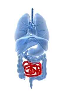
Background imageDigestive System Collection: X-ray image of internal organs with small intestine highlighted in red

X-ray image of internal organs with small intestine highlighted in red

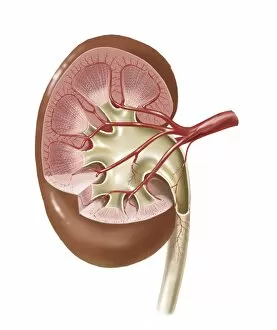
Background imageDigestive System Collection: Human kidney with artery

Human kidney with artery

Background imageDigestive System Collection: Cutaway view of the human kidney and adrenal gland

Cutaway view of the human kidney and adrenal gland

Background imageDigestive System Collection: Anatomy of human digestive system

Anatomy of human digestive system

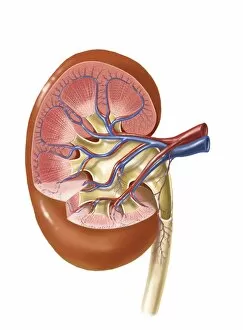
Background imageDigestive System Collection: Interior detail of human kidney

Interior detail of human kidney

Background imageDigestive System Collection: Human skeleton showing kidney, stomach and intestines

Human skeleton showing kidney, stomach and intestines
Human skeleton showing kidney, stomach, intestines and nervous system

Background imageDigestive System Collection: Medical illustration showing the human digestive system

Medical illustration showing the human digestive system: liver, stomach, large intestine, small intestine

Background imageDigestive System Collection: Anatomy of gall bladder ganglion

Anatomy of gall bladder ganglion

Background imageDigestive System Collection: Microscopic cross section view of the small intestine

Microscopic cross section view of the small intestine. TheAsmall intestineAis a long, highly convoluted tube in the digestive system that absorbs about 90% of the nutrients from the food we eat

Background imageDigestive System Collection: Front view of human digestive system with lungs

Front view of human digestive system with lungs

Background imageDigestive System Collection: X-ray image of internal organs with lungs highlighted in red

X-ray image of internal organs with lungs highlighted in red

Background imageDigestive System Collection: Urine production in human kidney

Urine production in human kidney

Background imageDigestive System Collection: Human kidney

Human kidney

Background imageDigestive System Collection: Medical illustration of female skeleton with veins and arteries

Medical illustration of female skeleton with veins and arteries

Background imageDigestive System Collection: Human body and digestive system, perspective view

Human body and digestive system, perspective view

Background imageDigestive System Collection: Human skeletal system with organs of the digestive system visible

Human skeletal system with organs of the digestive system visible

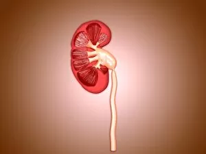
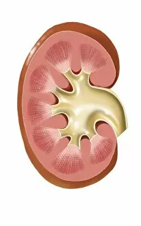
Background imageDigestive System Collection: Cross-section of human kidney

Cross-section of human kidney

Background imageDigestive System Collection: Illustration of diverticulosis in the colon

Illustration of diverticulosis in the colon. Diverticulitis is a common digestive disease which involves the formation of pouches (diverticula) within the bowel wall
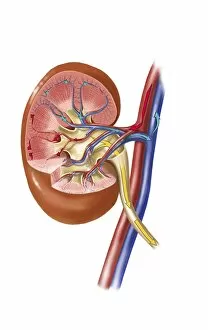
Background imageDigestive System Collection: Polycystic kidney

Polycystic kidney

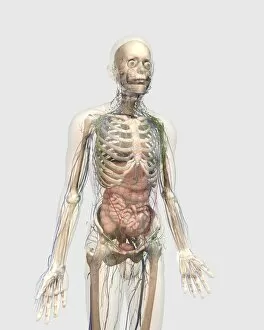
Background imageDigestive System Collection: Human body with internal organs, lymphatic system and circulatory system

Human body with internal organs, lymphatic system and circulatory system
Transparent human body with internal organs, lymphatic system and circulatory system

Background imageDigestive System Collection: Ureterovesical junction (UVJ) in the kidney

Ureterovesical junction (UVJ) in the kidney. This is the point where the ureters from the kidney connect to the bladder

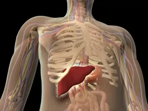
Background imageDigestive System Collection: Transparent view of human torso showing liver

Transparent view of human torso showing liver

Background imageDigestive System Collection: Front view of human digestive system

Front view of human digestive system

Background imageDigestive System Collection: Medical illustration of the human digestive system

Medical illustration of the human digestive system; salivary glands, esophagus, oral cavity


All Professionally Made to Order for Quick Shipping

The digestive system is a complex network of organs and processes that allows our bodies to break down food and absorb nutrients. From the gastrointestinal nematodes that inhabit our intestines to the medical illustration of an appendix with appendicitis, every component plays a vital role in this intricate system. Bee anatomy, depicted through artwork, showcases how these tiny creatures have their own unique digestive systems. Meanwhile, a scanning electron microscope (SEM) image reveals the Salmonella bacteria responsible for causing foodborne illnesses. Another SEM image focuses on the small intestine's surface, highlighting its crucial function in nutrient absorption. Similarly, we see the gallbladder's surface under SEM examination, emphasizing its role in storing bile produced by the liver. C. Elegans worms are also featured here as they provide valuable insights into understanding human digestion at a microscopic level. A light micrograph captures their intricate structure and behavior within this fascinating ecosystem. Moving beyond microscopic organisms, we encounter a muskrat sitting on the shore of a pond indulging in grass consumption—an example of herbivorous digestion within nature's cycle. An X-ray image provides us with an inside look at an inflamed appendix—a condition known as appendicitis—underscoring both its importance and potential health risks if left untreated. Lastly, artwork showcasing pancreas anatomy reminds us of this organ's significant contribution to digestion through hormone production such as insulin regulation.