Fraud Blocker Skip to main content

Digestive System Collection (page 35)

The digestive system is a complex network of organs and processes that allows our bodies to break down food and absorb nutrients

Background imageDigestive System Collection: Abdominal organs, anatomical artwork

Abdominal organs, anatomical artwork. The organs are shown within the surrounding bones, including the pelvis (bottom) and the rib-cage (top)

Background imageDigestive System Collection: Intestinal endocrine cell, TEM

Intestinal endocrine cell, TEM
Intestinal endocrine cell, coloured transmission electron micrograph (TEM). This is a type 1 intestinal endocrine cell. The granules (red) contain serotonin

Background imageDigestive System Collection: Bacterial diarrhoea, artwork

Bacterial diarrhoea, artwork
Bacterial diarrhoea. Computer artwork of Escherichia coli bacteria inside the intestines. The bacteria are magnified in size. Diarrhoea is loose, liquid bowel movements

Background imageDigestive System Collection: Helicobacter bilis bacteria, SEM

Helicobacter bilis bacteria, SEM
Helicobacter bilis bacteria, coloured field emission scanning electron micrograph (FESEM). H. bilis is a Gram-negative spiral-shaped bacterium found in the intestines of rodents, dogs and cats

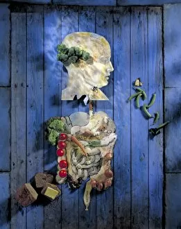
Background imageDigestive System Collection: Organic food, conceptual image

Organic food, conceptual image. Cut-out diagram of a mans head and digestive system, attached to a wooden background. Fruit and vegetables, and plastic sheep and pigs

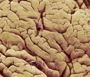
Background imageDigestive System Collection: Snake intestinal villi, SEM

Snake intestinal villi, SEM
Snake intestine. Coloured scanning electron micrograph (SEM) of villi in the intestine of a snake. The villi (folds) in the surface of the intestine are short and covered in cuboidal mucosal cells

Background imageDigestive System Collection: Snake intestine, SEM

Snake intestine, SEM
Snake intestine. Coloured scanning electron micrograph (SEM) of a freeze-fracture through the intestine of a snake. The mucosa (brown)

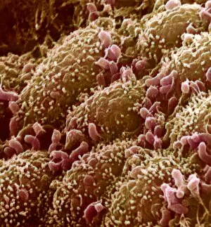
Background imageDigestive System Collection: Intestinal lining, SEM

Intestinal lining, SEM
Intestinal lining. Coloured scanning electron micrograph (SEM) of villi from the small intestine. Villi are finger-like projections that increase the surface area available for the absorption of

Background imageDigestive System Collection: Intestinal bacteria, SEM

Intestinal bacteria, SEM
Intestinal bacteria. Coloured scanning electron micrograph (SEM) of bacteria (green) on the surface of the duodenum. The duodenum is the first part of the small intestine

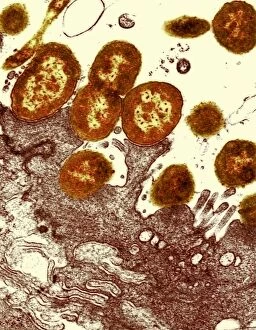
Background imageDigestive System Collection: Intestinal bacteria, TEM

Intestinal bacteria, TEM
Intestinal bacteria, coloured transmission electron micrograph (TEM). The TEM shows a section through the bacteria (red/yellow) and the wall of the intestines (bottom)

Background imageDigestive System Collection: Intestinal smooth muscle cells, TEM

Intestinal smooth muscle cells, TEM
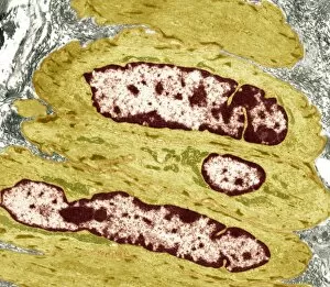
Intestinal smooth muscle cells. Coloured transmission electron micrograph (TEM) of a section through several smooth muscle cells from the intestines

Background imageDigestive System Collection: Pancreas acinus, SEM

Pancreas acinus, SEM
Pancreas acinus. Coloured scanning electron micrograph (SEM) of a freeze-fracture through an acinus (yellow) in the pancreas. An acinus is a collection of glandular epithelial cells

Background imageDigestive System Collection: Stomach pylorus glands, light micrograph

Stomach pylorus glands, light micrograph
Stomach pylorus glands. Light micrograph of a section through glands (pink) in the pylorus region of the stomach. Pyloric glands contain mucus cells and G cells that secrete gastrin

Background imageDigestive System Collection: Norwalk viral infection, artwork

Norwalk viral infection, artwork
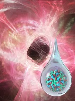
Norwalk viral infection. Conceptual computer artwork of infection of the gut (pink) by the norwalk virus, seen in a drop of water at lower right


All Professionally Made to Order for Quick Shipping

The digestive system is a complex network of organs and processes that allows our bodies to break down food and absorb nutrients. From the gastrointestinal nematodes that inhabit our intestines to the medical illustration of an appendix with appendicitis, every component plays a vital role in this intricate system. Bee anatomy, depicted through artwork, showcases how these tiny creatures have their own unique digestive systems. Meanwhile, a scanning electron microscope (SEM) image reveals the Salmonella bacteria responsible for causing foodborne illnesses. Another SEM image focuses on the small intestine's surface, highlighting its crucial function in nutrient absorption. Similarly, we see the gallbladder's surface under SEM examination, emphasizing its role in storing bile produced by the liver. C. Elegans worms are also featured here as they provide valuable insights into understanding human digestion at a microscopic level. A light micrograph captures their intricate structure and behavior within this fascinating ecosystem. Moving beyond microscopic organisms, we encounter a muskrat sitting on the shore of a pond indulging in grass consumption—an example of herbivorous digestion within nature's cycle. An X-ray image provides us with an inside look at an inflamed appendix—a condition known as appendicitis—underscoring both its importance and potential health risks if left untreated. Lastly, artwork showcasing pancreas anatomy reminds us of this organ's significant contribution to digestion through hormone production such as insulin regulation.