
Developmental Collection
"Unveiling the Beauty of Developmental Stages: From Reading Girl to Tadpoles and Beyond" In this captivating collection of images


All Professionally Made to Order for Quick Shipping
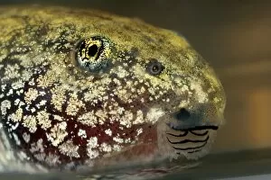
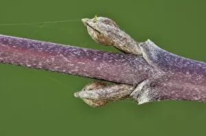
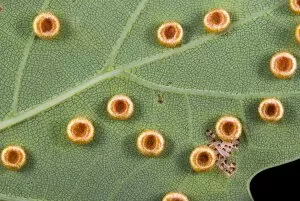
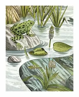
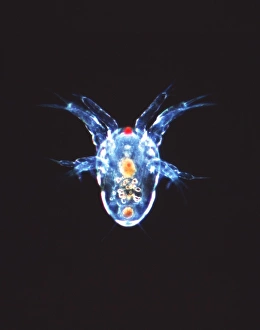
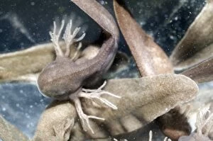
"Unveiling the Beauty of Developmental Stages: From Reading Girl to Tadpoles and Beyond" In this captivating collection of images, we embark on a visual journey through various developmental stages that showcase the marvels of life's progression. Starting with "Reading Girl, " a delicate pen and ink drawing on paper, we witness the early stages of intellectual growth as she immerses herself in the world of books. The innocence and curiosity radiating from her eyes hint at the potential for knowledge yet to be discovered. Moving forward, "Children at Breakfast" captures a moment frozen in time, painted meticulously with oil on canvas. Here, we witness not only physical development but also social interaction among young ones as they gather around a table filled with nourishment, and is an ode to childhood camaraderie and shared experiences. Shifting our focus towards nature's wonders, we encounter "Larvae of the Sawfly, " showcasing an insect's transformative journey from its earliest stage. This intricate depiction reminds us that even seemingly insignificant creatures undergo profound changes before revealing their true beauty. Our exploration takes an unexpected turn as X-ray images reveal "Melorheostosis" - abnormal bone growth affecting knees. These haunting visuals serve as reminders that development can sometimes take unpredictable paths, presenting challenges along the way. Returning to nature's realm once more, we encounter another caterpillar head – symbolic of metamorphosis itself – reminding us that change is constant and necessary for growth. Alongside it are developing tadpoles swimming gracefully in their aquatic habitat; they embody resilience amidst transformation. Lastly, our attention turns to medical scans depicting conditions such as spina bifida - highlighting both fragility and strength within human development. These powerful images remind us how crucial it is to understand and support individuals navigating unique journeys. Through this diverse collection spanning artistry and science alike, one common thread emerges: developmental processes shape all living beings profoundly.