Fraud Blocker Skip to main content

Cryo Em Collection

"Cryo-EM: Unlocking the Secrets of Microscopic Worlds" In the realm of scientific exploration

Background imageCryo Em Collection: Murine norovirus with antibody fragments

Murine norovirus with antibody fragments
Murine norovirus (MNV) with antibody fragments, computer model. This image was created using molecular modelling software and data from cryo- electron microscopy

Background imageCryo Em Collection: Cucumber necrosis virus, computer model

Cucumber necrosis virus, computer model
Cucumber necrosis virus (CNV), computer model. This image was created using molecular modelling software and data from cryo-electron microscopy

Background imageCryo Em Collection: Human rhinovirus, computer model

Human rhinovirus, computer model
Human rhinovirus (HRV) with intercellular adhesion molecule 1 (ICAM-1), computer model. This image was created using UCSF Chimera molecular modelling software and data from cryo-electron microscopy

Background imageCryo Em Collection: Ribgrass mosaic virus, computer model

Ribgrass mosaic virus, computer model
Ribgrass mosaic virus (RMV), computer model. This image was created using UCSF Chimera molecular modelling software and fibre X-ray diffraction

Background imageCryo Em Collection: Human rhinovirus with antibodies

Human rhinovirus with antibodies
Human rhinovirus (HRV) with antibody fragments, computer model. This image was created using molecular modelling software and data from cryo- electron microscopy

Background imageCryo Em Collection: Cucumber mosaic virus with antibodies

Cucumber mosaic virus with antibodies
Cucumber mosaic virus (CMV) with antibody fragments attached, computer model. This image was created using molecular modelling software and data from cryo-electron microscopy

Background imageCryo Em Collection: Bacteriophage phi29, computer model

Bacteriophage phi29, computer model
Bacteriophage phi29. Cross section computer model of phi29 created using UCSF Chimera molecular modelling software and data from cryo-electron microscopy

Background imageCryo Em Collection: Murine norovirus, computer model

Murine norovirus, computer model
Murine norovirus (MNV), computer model. This image was created using molecular modelling software and data from cryo-electron microscopy

Background imageCryo Em Collection: Simian immunodeficiency virus (SIV)

Simian immunodeficiency virus (SIV), computer model. Cross section of SIV created using 3-D molecular modelling software and data from cryo- electron microscopy

Background imageCryo Em Collection: Sindbis virus, computer model

Sindbis virus, computer model
Sindbis virus. Computer model of sindbis virus created using UCSF Chimera molecular modelling software and data from cryo-electron microscopy. It shows the outer glycoprotein shell of the virus

Background imageCryo Em Collection: Semliki Forest virus, computer model

Semliki Forest virus, computer model
Semliki Forest virus (SFV), computer model. This image was created using UCSF Chimera molecular modelling software and data from cryo-electron microscopy


All Professionally Made to Order for Quick Shipping

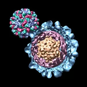
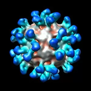
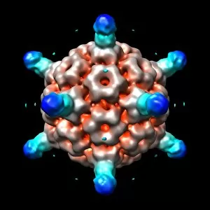
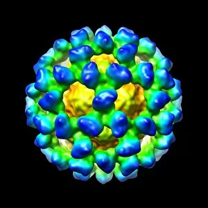
"Cryo-EM: Unlocking the Secrets of Microscopic Worlds" In the realm of scientific exploration, Cryo-Electron Microscopy (Cryo-EM) has emerged as a groundbreaking technique, revolutionizing our understanding of microscopic structures. With its ability to capture high-resolution images of biological specimens in their native state, Cryo-EM has paved the way for remarkable discoveries. One such revelation involves Bacteriophage phi29, where Cryo-EM enabled scientists to visualize this tiny virus with unprecedented detail. By combining experimental data and computer models, researchers gained insights into its intricate architecture and mechanisms that drive infection. Simian immunodeficiency virus (SIV), a close relative of HIV, also fell under the scrutiny of Cryo-EM. This powerful imaging technique allowed scientists to study SIV's structural features at an atomic level, providing crucial information for potential antiviral strategies. The application of Cryo-EM extended beyond viruses alone. Murine norovirus with antibody fragments became another subject for investigation. Through cryogenic preservation and electron microscopy techniques, researchers were able to observe how these antibodies interacted with the virus particles - knowledge that could aid in designing effective therapeutics against norovirus infections. Expanding on previous successes with Bacteriophage phi29 and Cucumber necrosis virus using computer models; Cryo-EM offered new avenues for studying other viruses like Human rhinovirus and Ribgrass mosaic virus. These computer-generated models provided valuable insights into viral structure-function relationships while complementing experimental findings obtained through cryogenic imaging. With further advancements in technology came breakthroughs involving Human rhinovirus coupled with antibodies. By visualizing these interactions using Cryo-EM techniques, researchers gained critical information about how antibodies neutralize viral threats within our bodies – knowledge instrumental in developing targeted treatments against respiratory illnesses caused by rhinoviruses. Bacteriophage P22 joined the ranks as another virus studied using Cryo-EM and computer models.