
Cone Cell Collection
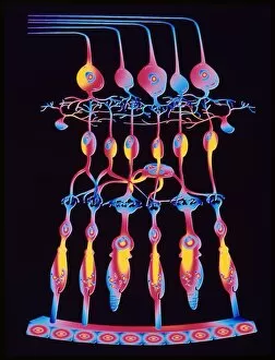
Caption: Unveiling the Marvels of Cone Cells in the Eye's Retina In this captivating collection of scanning electron microscope (SEM) images


All Professionally Made to Order for Quick Shipping
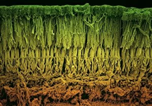
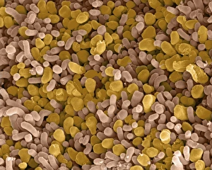
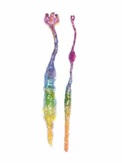
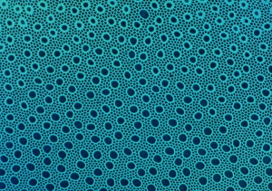
Caption: Unveiling the Marvels of Cone Cells in the Eye's Retina In this captivating collection of scanning electron microscope (SEM) images, we delve into the intricate world of cone cells - one of the key components responsible for our visual perception. The first image, SEM C014 / 4866, offers a mesmerizing close-up view of rod and cone cells within the eye's retina. These specialized photoreceptor cells play a vital role in converting light into electrical signals that are then transmitted to our brain. Moving on to SEM C014 / 4864, we witness another stunning portrayal showcasing rod and cone cells working harmoniously together. Each cell type possesses unique characteristics; rods excel at detecting dim light levels while cones specialize in perceiving color and fine details. Zooming further into the realm of these remarkable structures, we encounter an exquisite SEM image displaying both rod and cone cells within the retina (Eye retina F008 / 0713). This snapshot highlights their distinct shapes and arrangements, emphasizing their crucial roles in shaping our visual experiences. As we explore subsequent images such as Eye retina F008 / 0719 or Eye retina F008 / 0712, it becomes evident how densely packed these delicate photoreceptor cells are within our retinas. Their sheer abundance ensures optimal vision by capturing every nuance of light that enters our eyes. Eye retina F008 / 0715 showcases a closer look at individual cone cells with their characteristic tapered shape. These specialized receptors enable us to perceive vibrant colors across different wavelengths - from fiery reds to soothing blues - enriching our daily encounters with the world around us. The remaining snapshots continue to unveil more intricacies hidden within this microscopic landscape. From Eye retina F008 / 0717 to Eye retina F008 /0720, each image provides glimpses into various regions where these extraordinary cone cells reside – forming an awe-inspiring mosaic dedicated solely to our visual perception.