
Cell Membrane Collection
The cell membrane, also known as the plasma membrane, is a fascinating structure that plays a crucial role in maintaining the integrity and functionality of cells


All Professionally Made to Order for Quick Shipping
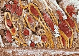
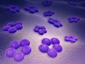
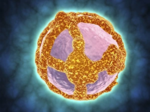
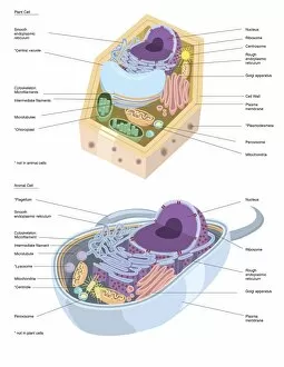
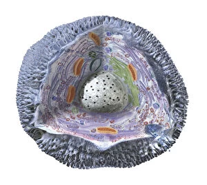
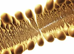
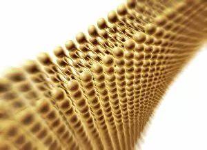
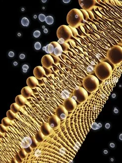
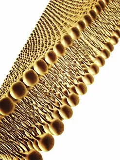
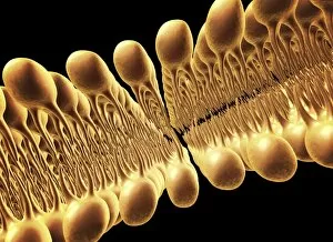
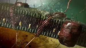
The cell membrane, also known as the plasma membrane, is a fascinating structure that plays a crucial role in maintaining the integrity and functionality of cells. This intricate artwork (C013 / 7467) beautifully captures its complexity. One important aspect of the cell membrane is its ability to interact with various molecules and substances. The artwork depicting cannabinoid receptor binding highlights this interaction, showcasing how these receptors bind to specific molecules within the membrane. Another captivating image shows AIDS viruses budding from a cell under a transmission electron microscope (TEM). This powerful visual reminds us of the devastating impact that certain viruses can have on our immune system, such as the simian immunodeficiency virus (SIV). While many cellular structures are enclosed by membranes, mitochondria stand out due to their unique characteristics. A scanning electron microscope (SEM) image showcases their distinctive shape and structure, reminding us of their vital role in energy production within cells. Understanding animal cell structure is essential for comprehending cellular functions. Microscopic views provide valuable insights into these complex structures and help scientists unravel their mysteries. Artwork F007 / 1477 depicts the lipid bilayer composition of the cell membrane. This conceptual representation emphasizes how lipids arrange themselves to form a barrier between the intracellular and extracellular environments. Proteins embedded within the cell membrane facilitate numerous processes by forming channels for ion transport. The MscL ion channel protein structure gives us an insight into one such protein's architecture responsible for regulating ion flow across membranes. Once again, SEM imaging allows us to appreciate mitochondria's beauty while highlighting their importance in cellular function – this time through another stunning depiction. Microscopic views offer glimpses into diverse aspects of cellular life – from overall morphology to intricate details like surface receptors or even radiolarians with skeletal frames captured conceptually - illustrating nature's incredible diversity at microscopic levels.