Fraud Blocker Skip to main content

Brain Collection (page 3)

"The intricate map of the brain: Exploring its motor and sensory pathways" Delving into the depths of our mind, we uncover the wonders of the brain's complex anatomy

Background imageBrain Collection: Brain

Brain. Computer artwork of a human brain inside the skull

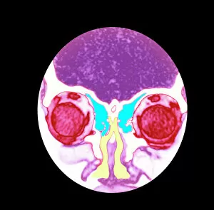
Background imageBrain Collection: Nose and sinuses, CT scan

Nose and sinuses, CT scan
Nose anatomy and sinuses. Coloured computed tomography (CT) scan of a frontal view through the nose of a 31 year old woman

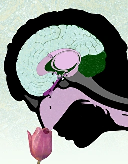
Background imageBrain Collection: Smell pathway

Smell pathway. Artwork of the pathway involved in the sensation of smelling a flower. The perception of smell starts in the lining (mucosa) of the nose (1)

Background imageBrain Collection: Astrocyte nerve cell

Astrocyte nerve cell. Fluorescent light micrograph of an astrocyte cell from a human brain. Intermediate filaments (IFs), part of the cells cytoskeleton, have been dyed green

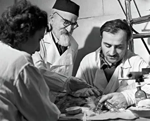
Background imageBrain Collection: Beritashvili and students, Tbilisi, 1962

Beritashvili and students, Tbilisi, 1962
Ivan Beritashvili (1885-1974, centre), Soviet physiologist, instructing students who are studying the brain of a laboratory animal

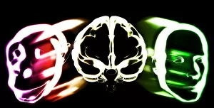
Background imageBrain Collection: Primate brain evolution

Primate brain evolution. Conceptual computer artwork of a brain (centre) with a monkey head (left) and a human head (right), representing the evolution of primate brains

Background imageBrain Collection: Serotonin neurotransmitter molecule

Serotonin neurotransmitter molecule
Serotonin molecule. Computer artwork of a molecule of serotonin (5-hydroxytryptamine, C10.H12.N2.O), a neurotransmitter. Atoms (spheres) are colour- coded: carbon (black), hydrogen (white)

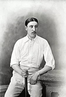
Background imageBrain Collection: Cricketer, Brain

Cricketer, Brain
J.H. Brain Cricketer - Gloucestershire & Glamorgan

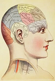
Background imageBrain Collection: A Phrenological Map Of The Human Brain. From Virtues Household Physician, Published London 1924

A Phrenological Map Of The Human Brain. From Virtues Household Physician, Published London 1924

Background imageBrain Collection: Front view of the female anatomy hilighting the endocrine system

Front view of the female anatomy hilighting the endocrine system
Brain, Anatomy, Diagram, Heart, Illustration, Bone, Shoulder, Medical, Uterus, Biology, Pelvis, Femur, See Through, Vertical, Artwork, Female Likeness, Color Image, Front View

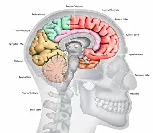
Background imageBrain Collection: Human brain, illustration

Human brain, illustration
Illustration of a cross-section of the brain showing the various lobes. The lobes are shown in different colours - red (frontal), green (parietal), yellow (occipital), orange (temporal)

Background imageBrain Collection: Advertisement for Harness' Electropathic Belt (engraving)

Advertisement for Harness' Electropathic Belt (engraving)
8631622 Advertisement for Harness' Electropathic Belt (engraving) by English School, (19th century); Private Collection; (add.info.: Advertisement for Harness' Electropathic Belt)

Background imageBrain Collection: Map of nerve bundles in the human brain by Paul Broca

Map of nerve bundles in the human brain by Paul Broca
5309460 Map of nerve bundles in the human brain by Paul Broca; (add.info.: Map of nerve bundles in the human brain by Paul Broca)

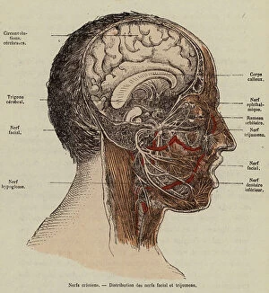
Background imageBrain Collection: Nerves of the face (coloured engraving)

Nerves of the face (coloured engraving)
8655732 Nerves of the face (coloured engraving) by French School, (19th century) (after); Private Collection; (add.info.: Nerves of the face)

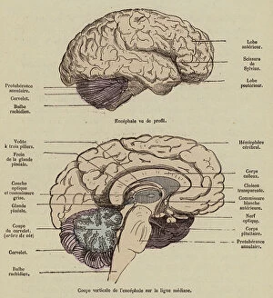
Background imageBrain Collection: Brain viewed in profile and cross-section (coloured engraving)

Brain viewed in profile and cross-section (coloured engraving)
8655728 Brain viewed in profile and cross-section (coloured engraving) by French School, (19th century) (after); Private Collection; (add.info.: Brain viewed in profile and cross-section)

Background imageBrain Collection: Connection of the sympathetic trunk with the vagus nerve and the spinal nerves (coloured engraving)

Connection of the sympathetic trunk with the vagus nerve and the spinal nerves (coloured engraving)
8655724 Connection of the sympathetic trunk with the vagus nerve and the spinal nerves (coloured engraving) by French School

Background imageBrain Collection: Olfactory and maxillary nerves. Lithograph by Davesne, based on a drawing by Leveille

Olfactory and maxillary nerves. Lithograph by Davesne, based on a drawing by Leveille
FLO4672503 Olfactory and maxillary nerves. Lithograph by Davesne, based on a drawing by Leveille, in Petity Atlas complet d'Anatomie descriptive du Corps Humain, by Dr

Background imageBrain Collection: Saphene vein and medial plantar nerves. Lithograph of Corbie, based on a drawing by Leveille

Saphene vein and medial plantar nerves. Lithograph of Corbie, based on a drawing by Leveille
FLO4672470 Saphene vein and medial plantar nerves. Lithograph of Corbie, based on a drawing by Leveille, in Petity Atlas complet d'Anatomie descriptive du Corps Humain, by Dr

Background imageBrain Collection: Brain sections, cerebellum and ventricles. Lithograph by Davesne, based on a drawing by Leveille

Brain sections, cerebellum and ventricles. Lithograph by Davesne, based on a drawing by Leveille
FLO4673145 Brain sections, cerebellum and ventricles. Lithograph by Davesne, based on a drawing by Leveille, in Petity Atlas complet d'Anatomie descriptive du Corps Humain, by Dr

Background imageBrain Collection: Cross sections of the brain. Lithograph of Corbie, based on a drawing by Leveille

Cross sections of the brain. Lithograph of Corbie, based on a drawing by Leveille
FLO4673108 Cross sections of the brain. Lithograph of Corbie, based on a drawing by Leveille, in Petity Atlas complet d'Anatomie descriptive du Corps Humain, by Dr

Background imageBrain Collection: The circulatory system of the brain. Lithograph of Choubard, based on a drawing by Leveille

The circulatory system of the brain. Lithograph of Choubard, based on a drawing by Leveille
FLO4672884 The circulatory system of the brain. Lithograph of Choubard, based on a drawing by Leveille, in Petity Atlas complet d'Anatomie descriptive du Corps Humain, by Dr

Background imageBrain Collection: Phrenological Chart

Phrenological Chart
2626135 Phrenological Chart; (add.info.: Phrenological chart of 1859. phrenology was founded in the early 19th century by Franz Josef Gall (1758-1828)

Background imageBrain Collection: Interior of the brain and cerebellum. Copperplate engraving by Robert Benard after an illustration

Interior of the brain and cerebellum. Copperplate engraving by Robert Benard after an illustration by Albrecht von
FLO4611652 Interior of the brain and cerebellum. Copperplate engraving by Robert Benard after an illustration by Albrecht von Haller from Denis Diderot's Encyclopedia, Pellet, Geneva

Background imageBrain Collection: Sympathic nervous system in the thoracic, lumbar and sacral plexus. Lithograph by Davesne

Sympathic nervous system in the thoracic, lumbar and sacral plexus. Lithograph by Davesne
FLO4672529 Sympathic nervous system in the thoracic, lumbar and sacral plexus. Lithograph by Davesne, based on a drawing by Leveille

Background imageBrain Collection: Sympathic nervous system in the cardiac plexus. Lithograph by Davesne

Sympathic nervous system in the cardiac plexus. Lithograph by Davesne, based on a drawing by Leveille
FLO4672521 Sympathic nervous system in the cardiac plexus. Lithograph by Davesne, based on a drawing by Leveille, in Petiure Atlas complet d'Anatomie descriptive du Corps Humain, by Dr

Background imageBrain Collection: Nervous system of the back. Lithograph of Corbie, based on a drawing by Leveille

Nervous system of the back. Lithograph of Corbie, based on a drawing by Leveille
FLO4672457 Nervous system of the back. Lithograph of Corbie, based on a drawing by Leveille, in Petity Atlas complet d'Anatomie descriptive du Corps Humain, by Dr

Background imageBrain Collection: Intercostal nerves. Lithograph of Corbie, based on a drawing by Leveille

Intercostal nerves. Lithograph of Corbie, based on a drawing by Leveille, in Petity Atlas complet d'Anatomie
FLO4672415 Intercostal nerves. Lithograph of Corbie, based on a drawing by Leveille, in Petity Atlas complet d'Anatomie descriptive du Corps Humain, by Dr

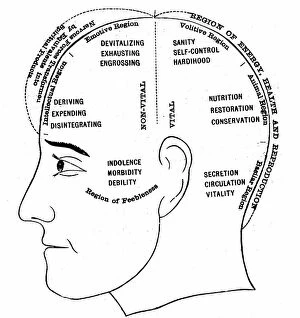
Background imageBrain Collection: An engraving depicting a diagram showing the supposed functions of various areas of the brain

An engraving depicting a diagram showing the supposed functions of various areas of the brain, 19th century
5311364 An engraving depicting a diagram showing the supposed functions of various areas of the brain, 19th century; (add.info.: Date: 1886); Universal History Archive/UIG.

Background imageBrain Collection: Brain, pons, spinal bulb, pineal gland etc... Lithograph by Davesne, based on a drawing by Leveille

Brain, pons, spinal bulb, pineal gland etc... Lithograph by Davesne, based on a drawing by Leveille
FLO4673157 Brain, pons, spinal bulb, pineal gland etc... Lithograph by Davesne, based on a drawing by Leveille, in Petity Atlas complet d'Anatomie descriptive du Corps Humain, by Dr

Background imageBrain Collection: Section through the brain. Lithograph by Davesne, based on a drawing by Leveille

Section through the brain. Lithograph by Davesne, based on a drawing by Leveille
FLO4673112 Section through the brain. Lithograph by Davesne, based on a drawing by Leveille, in Petity Atlas complet d'Anatomie descriptive du Corps Humain, by Dr

Background imageBrain Collection: Rene Descartes' idea of vision

Rene Descartes' idea of vision
5310106 Rene Descartes' idea of vision; (add.info.: Diagram illustrating Rene Descartes' idea of vision, showing the function of the eye, optic nerve, and brain)

Background imageBrain Collection: Nerves damaged by war (litho)

Nerves damaged by war (litho)
8664192 Nerves damaged by war (litho) by English School, (20th century); Private Collection; (add.info.: Nerves damaged by war)

Background imageBrain Collection: 8623776

8623776

Background imageBrain Collection: An engraving depicting Descartes' drawing of the human brain, 17th century

An engraving depicting Descartes' drawing of the human brain, 17th century
5311365 An engraving depicting Descartes' drawing of the human brain, 17th century; Universal History Archive/UIG.

Background imageBrain Collection: Phrenology (anatomical), illustration by F. E. Guerin. From Dictionnaire pittoresque d'histoire

Phrenology (anatomical), illustration by F. E. Guerin. From Dictionnaire pittoresque d'histoire naturelle et des
5305673 Phrenology (anatomical), illustration by F. E. Guerin. From Dictionnaire pittoresque d'histoire naturelle et des phenomenes de la nature-1833/1834; (add.info.: Phrenology (anatomical)

Background imageBrain Collection: Inca Crane proving the practice of surgical operation on the human brain during prehistory in Peru

Inca Crane proving the practice of surgical operation on the human brain during prehistory in Peru Colour engraving of
NWI4900256 Inca Crane proving the practice of surgical operation on the human brain during prehistory in Peru Colour engraving of the 19th century; (add.info)

Background imageBrain Collection: Brain sections. Lithograph by Davesne, based on a drawing by Leveille

Brain sections. Lithograph by Davesne, based on a drawing by Leveille, in Petity Atlas complet d'Anatomie
FLO4673204 Brain sections. Lithograph by Davesne, based on a drawing by Leveille, in Petity Atlas complet d'Anatomie descriptive du Corps Humain, by Dr

Background imageBrain Collection: Ophthalmic and maxillary nerves of the fifth cranial nerve. Lithograph by Davesne

Ophthalmic and maxillary nerves of the fifth cranial nerve. Lithograph by Davesne, based on a drawing by Leveille
FLO4672496 Ophthalmic and maxillary nerves of the fifth cranial nerve. Lithograph by Davesne, based on a drawing by Leveille, in Petity Atlas complet d'Anatomie descriptive du Corps Humain

Background imageBrain Collection: Lumbar plexus and crural nerve. Lithograph of Corbie, based on a drawing by Leveille

Lumbar plexus and crural nerve. Lithograph of Corbie, based on a drawing by Leveille
FLO4672455 Lumbar plexus and crural nerve. Lithograph of Corbie, based on a drawing by Leveille, in Petity Atlas complet d'Anatomie descriptive du Corps Humain, by Dr

Background imageBrain Collection: Asian tapeworm, Bothriocephalus acheilognathi (Botryocephalus latus), freshwater fish parasite

Asian tapeworm, Bothriocephalus acheilognathi (Botryocephalus latus), freshwater fish parasite
FLO4720090 Asian tapeworm, Bothriocephalus acheilognathi (Botryocephalus latus), freshwater fish parasite, and Coenurus cerebralis, larva of the tapeworm Multiceps multiceps

Background imageBrain Collection: Ernst Heinrich Weber, 1820

Ernst Heinrich Weber, 1820
1576985 Ernst Heinrich Weber, 1820; (add.info.: Ernst Heinrich Weber (June 24, 1795 - January 26, 1878) was a German physician

Background imageBrain Collection: Epilepsy, a subject suffering from a typical Grand Mal attack in which he has bitten his tongue

Epilepsy, a subject suffering from a typical Grand Mal attack in which he has bitten his tongue
527399 Epilepsy, a subject suffering from a typical Grand Mal attack in which he has bitten his tongue. In ancient times it was known as the Sacred Disease

Background imageBrain Collection: A woman's mind magnified (colour litho)

A woman's mind magnified (colour litho)
8635593 A woman's mind magnified (colour litho) by English School, (20th century); Private Collection; (add.info.: A woman's mind magnified)

Background imageBrain Collection: Nerves involved in the sense of taste (coloured engraving)

Nerves involved in the sense of taste (coloured engraving)
8655717 Nerves involved in the sense of taste (coloured engraving) by French School, (19th century) (after); Private Collection; (add.info.: Nerves involved in the sense of taste)

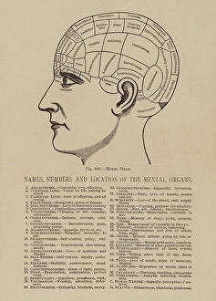
Background imageBrain Collection: Names, Numbers and Location of the Mental Organs (litho)

Names, Numbers and Location of the Mental Organs (litho)
8641361 Names, Numbers and Location of the Mental Organs (litho) by American School, (19th century); Private Collection; (add.info.: Names, Numbers and Location of the Mental Organs)

Background imageBrain Collection: Phrenological chart of an average girl's head (litho)

Phrenological chart of an average girl's head (litho)
8656500 Phrenological chart of an average girl's head (litho) by English School, (20th century); Private Collection; (add.info.: Phrenological chart of an average girl's head)

Background imageBrain Collection: Cross-section of the neck at the leval of the sixth cervical vertebra (coloured engraving)

Cross-section of the neck at the leval of the sixth cervical vertebra (coloured engraving)
8655734 Cross-section of the neck at the leval of the sixth cervical vertebra (coloured engraving) by French School, (19th century) (after); Private Collection; (add.info)

Background imageBrain Collection: Brain seen from below (coloured engraving)

Brain seen from below (coloured engraving)
8655729 Brain seen from below (coloured engraving) by French School, (19th century) (after); Private Collection; (add.info.: Brain seen from below)


All Professionally Made to Order for Quick Shipping

"The intricate map of the brain: Exploring its motor and sensory pathways" Delving into the depths of our mind, we uncover the wonders of the brain's complex anatomy. The Motor homunculus model reveals a distorted representation of our body, with certain regions magnified to depict their significance in movement control. Similarly, the Sensory homunculus showcases how different areas are dedicated to processing various sensations, emphasizing their importance in perceiving touch, pain, and temperature. Transporting us back in time, an engraving from 1895 unveils a vintage depiction anatomy. This historical artwork reminds us of the continuous quest for understanding this enigmatic organ that has captivated scientists for centuries. Zooming closer under a microscope lens, we witness the delicate beauty within cerebellum tissue through a light micrograph. Its intricate structure plays a crucial role in coordinating movements and maintaining balance. The Brain fibres captured by DTI MRI scan C017 / 7099 provide insight into its connectivity network. These fibers act as highways transmitting information between different regions - like bridges connecting distant lands. An inferior view offers another perspective on human brain anatomy. It unravels hidden complexities beneath our surface thoughts – showcasing structures responsible for memory formation, language processing, and emotional regulation. Artistic imagination breathes life into white matter fibres and brains through artwork C015 / 1930. This captivating portrayal merges science with creativity to portray both scientific accuracy and aesthetic appeal simultaneously. Venturing deeper within this mysterious realm is an exploration blood vessels using a stunning 3D angiogram C007 /1981. These intricate networks supply oxygen-rich blood to nourish every nook and cranny of this vital organ. Returning once again to explore motor and sensory functions intricately mapped out by homunculi models – these visual representations highlight how specific regions control precise movements or perceive distinct sensations throughout our body A masterpiece artwork captures Medulla oblongata in the brain, showcasing its vital role in controlling essential functions