Fraud Blocker Skip to main content

Brain Collection (page 2)

"The intricate map of the brain: Exploring its motor and sensory pathways" Delving into the depths of our mind, we uncover the wonders of the brain's complex anatomy

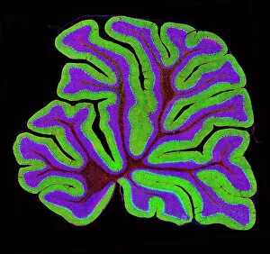
Background imageBrain Collection: Cerebellum structure, light micrograph

Cerebellum structure, light micrograph
Cerebellum structure. Fluorescent light micrograph of a section through the cerebellum of the brain. The cerebellum comprises three main layers

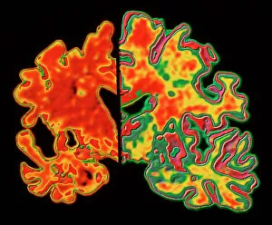
Background imageBrain Collection: Alzheimers brain

Alzheimers brain. Computer processed vertical (coronal) slice through the brain of an Alzheimer patient (at left) compared with a normal brain (at right)

Background imageBrain Collection: Brain protein research

Brain protein research. Computer artwork of a brain and coloured dots from a protein microarray. Protein microarrays can be used to follow protein interactions

Background imageBrain Collection: Brain anatomy, artwork

Brain anatomy, artwork
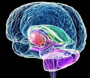
Brain anatomy, computer artwork. The cerebellum is purple the corpus callosum is green

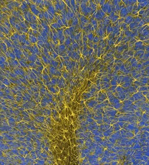
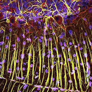
Background imageBrain Collection: Glial cells, confocal light micrograph

Glial cells, confocal light micrograph
Glial cells. Confocal light micrograph of glial cells from the cerebellum of the brain. Glial cells are nervous system cells that provide structural support and protection for neurons (nerve cells)

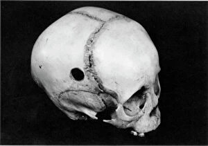
Background imageBrain Collection: TREPANNING: SKULL. Trepanned Pre-Columbian skull found in Peru

TREPANNING: SKULL. Trepanned Pre-Columbian skull found in Peru

Background imageBrain Collection: Ovaltine Advert

Ovaltine Advert
Advertisement for Ovaltine which " builds up brain, nerve and body"

Background imageBrain Collection: Surgery Cures Stupidity

Surgery Cures Stupidity
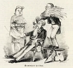
SATIRE:RESULTS OF ETHER - surgery as a cure-all: young mans stupidity cured by replacing straw head- stuffing with a brain!

Background imageBrain Collection: Brain anatomy, artwork

Brain anatomy, artwork
Brain anatomy, computer artwork. The cerebellum is purple the corpus callosum is green

Background imageBrain Collection: Brain anatomy, artwork

Brain anatomy, artwork
Brain anatomy, computer artwork. The cerebellum is purple the corpus callosum is green

Background imageBrain Collection: Cerebellum tissue, light micrograph

Cerebellum tissue, light micrograph
Cerebellum tissue. Confocal light micrograph of a section through the cerebellum of the brain showing two types of glial cells (support cells); astrocytes (star-shaped)

Background imageBrain Collection: WHAT THE BRAIN DOES

WHAT THE BRAIN DOES
What the brain does

Background imageBrain Collection: Side View of Brain / C19

Side View of Brain / C19
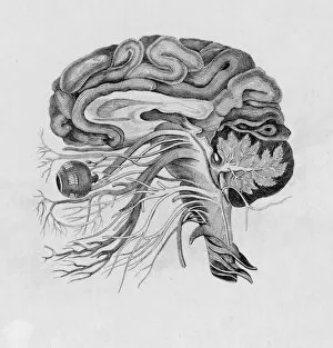
A side view

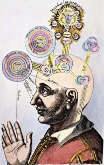
Background imageBrain Collection: English physician and Rosicrucian. Human mental abilities classified in terms of God

English physician and Rosicrucian. Human mental abilities classified in terms of God and the universe
ROBERT FLUDD (1574-1637). English physician and Rosicrucian. Human mental abilities classified in terms of God and the universe. Colored engraving from Fludds 17th century treatise, Utriusque Cosmi

Background imageBrain Collection: Startling Stories Scifi Magazine Cover with Science Island

Startling Stories Scifi Magazine Cover with Science Island
SCIENCE ISLAND (Eando Binder)A cold-blooded Scientific Napoleon with the Brain of a Genius and a Body of Metal threatens to Dominate Mankind Date: 1939

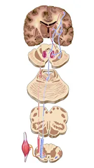
Background imageBrain Collection: Brain motor cortex pathways, artwork C016 / 6532

Brain motor cortex pathways, artwork C016 / 6532
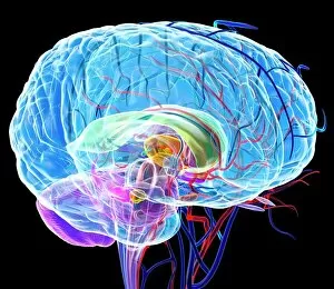
Brain motor cortex pathways. Artwork of a sectioned human brain, brainstem and spinal cord, showing neural pathways (red and blue) from the motor cortex of the brain

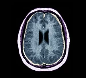
Background imageBrain Collection: Bacterial meningitis, MRI scan

Bacterial meningitis, MRI scan
Bacterial meningitis. Coloured magnetic resonance imaging (MRI) scan of an axial section through the brain of a 16-year-old patient

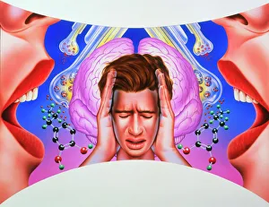
Background imageBrain Collection: Schizophrenia

Schizophrenia. Artwork of a man hearing non- existent womens voices. Auditory hallucinations are one of the most common symptoms of schizophrenia

Background imageBrain Collection: Brain neuron

Brain neuron. Computer reconstruction of a medium spiny neuron from the basal ganglia of the brain. Neurons (nerve cells) are responsible for passing information around the central nervous system

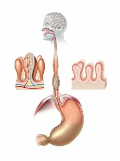
Background imageBrain Collection: Human digestive system, artwork

Human digestive system, artwork. At top, food from the mouth forms a bolus that is swallowed down the oesophagus. At centre, the bolus is about to enter the stomach (arrow)

Background imageBrain Collection: Cerebellum tissue, light micrograph

Cerebellum tissue, light micrograph
Cerebellum tissue. Confocal light micrograph of a section through the cerebellum of the brain. Purkinje cells, a type of neuron (nerve cell), are red

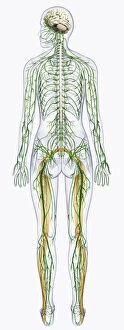
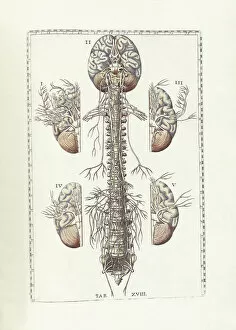
Background imageBrain Collection: Digital illustration of human nervous system connected to spinal cord and brain

Digital illustration of human nervous system connected to spinal cord and brain

Background imageBrain Collection: Digital illustration of Bocas area (purple), Wernickes area (orange)

Digital illustration of Bocas area (purple), Wernickes area (orange), Geschwinds territory (turquoise) and arcuate fasciculus (green) in human brain

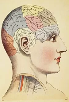
Background imageBrain Collection: A Phrenological Map Of The Human Brain

A Phrenological Map Of The Human Brain

Background imageBrain Collection: The human body with superimposed colored plates by Julien Bougle

The human body with superimposed colored plates by Julien Bougle
The human body with superimposed colored plates, by Julien Bougle, circa 1899

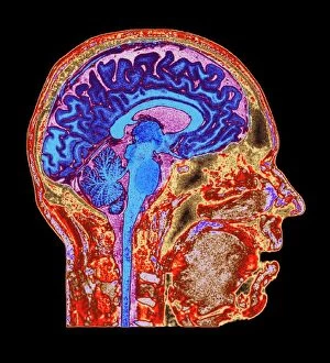
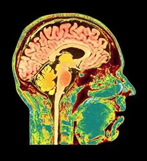
Background imageBrain Collection: Normal human brain, MRI scan C016 / 8845

Normal human brain, MRI scan C016 / 8845
Brain. Coloured magnetic resonance imaging (MRI) scan of a sagittal section through a patients head showing a healthy human brain

Background imageBrain Collection: Phantom pain after amputation, artwork

Phantom pain after amputation, artwork
Phantom pain after amputation. Artwork of a man experiencing phantom pain (red flash) from his amputated hand (left). This is caused by stimuli to the motor and pre-motor cortical areas of his brain

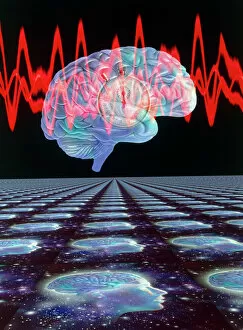
Background imageBrain Collection: Abstract artwork of human brain & EEG brainwaves

Abstract artwork of human brain & EEG brainwaves

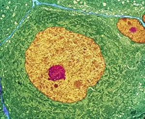
Background imageBrain Collection: Nerve cell, TEM

Nerve cell, TEM
Nerve cell. Coloured transmission electron micrograph (TEM) of a nerve cell body in cross- section. The cell has a large nucleus (yellow) and inner nucleolus (red)

Background imageBrain Collection: Decision-making hierarchy

Decision-making hierarchy
Decision making. Conceptual computer artwork of a hierarchy of decision makers, showing a lower level of 25 brains, which pass their decisions to a middle level of nine brains

Background imageBrain Collection: Healthy brain, MRI scans

Healthy brain, MRI scans
Healthy brain. Coloured magnetic resonance imaging (MRI) scans through the head of a 55 year old man, showing the brain sectioned in coronal (frontal) view

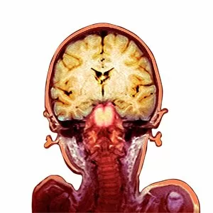
Background imageBrain Collection: Childs brain, MRI scan

Childs brain, MRI scan
Childs brain. Coloured magnetic resonance imaging (MRI) scan of an coronal (vertical) section through the head and brain of a healthy 3-year-old child, passing through the ears

Background imageBrain Collection: Dennis Brain Letter 1955

Dennis Brain Letter 1955
A letter from Dennis Brain, French horn player, to an admirer, enclosing his photograph

Background imageBrain Collection: The science of human anatomy by Bartholomeo Eustachi

The science of human anatomy by Bartholomeo Eustachi, depicting the shape, size and relative position of the organs of the human body. Circa 18th century

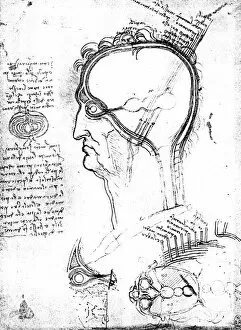
Background imageBrain Collection: Longitudinal section of an onion and sections of the human head

Longitudinal section of an onion and sections of the human head, eye and orbit: drawing by Leonardo da Vinci
LEONARDO: BRAIN, c1490. Longitudinal section of an onion and sections of the human head, eye and orbit: drawing by Leonardo da Vinci

Background imageBrain Collection: Artificial intelligence

Artificial intelligence. Conceptual computer artwork of a brain inside a computer, representing intelligent computers. The monitor screen shows a human eye

Background imageBrain Collection: George Wilhlem Friedrich Hegel, German philosopher

George Wilhlem Friedrich Hegel, German philosopher
George Wilhlem Friedrich Hegel (1770-1831), German idealist philosopher. His difficult philosophy, considered by many to be obscure, has great influence on Marxists, Positivists and Existentialists

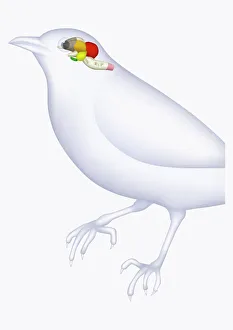
Background imageBrain Collection: Illustration of bird brain, including cerebrum, cerebellum, and medulla

Illustration of bird brain, including cerebrum, cerebellum, and medulla

Background imageBrain Collection: Digital illustration of direction of information in amygdala

Digital illustration of direction of information in amygdala

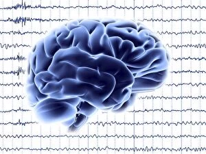
Background imageBrain Collection: Brain activity, artwork F006 / 4594

Brain activity, artwork F006 / 4594
Brain activity. Computer artwork of EEG (electroencephalogram) traces and a brain illustration. An EEG records the brains activity

Background imageBrain Collection: Normal human brain, MRI scan C016 / 8850

Normal human brain, MRI scan C016 / 8850
Brain. Coloured magnetic resonance imaging (MRI) scan of a sagittal section through a patients head showing a healthy human brain

Background imageBrain Collection: Purkinje nerve cell, TEM C014 / 0583

Purkinje nerve cell, TEM C014 / 0583
Purkinje nerve cell. Transmission electron micrograph (TEM) of a purkinje nerve cell (green) from the cerebellum of the brain, showing the cell body (centre) and its primary dendrite (cell process)

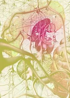
Background imageBrain Collection: Brain anatomy

Brain anatomy. Computer artwork of strands of nerve cells (neurons) in front of an exploded view of the brain seen from behind

Background imageBrain Collection: Brain

Brain. Computer artwork of a human brain inside the skull

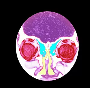
Background imageBrain Collection: Nose and sinuses, CT scan

Nose and sinuses, CT scan
Nose anatomy and sinuses. Coloured computed tomography (CT) scan of a frontal view through the nose of a 31 year old woman


All Professionally Made to Order for Quick Shipping

"The intricate map of the brain: Exploring its motor and sensory pathways" Delving into the depths of our mind, we uncover the wonders of the brain's complex anatomy. The Motor homunculus model reveals a distorted representation of our body, with certain regions magnified to depict their significance in movement control. Similarly, the Sensory homunculus showcases how different areas are dedicated to processing various sensations, emphasizing their importance in perceiving touch, pain, and temperature. Transporting us back in time, an engraving from 1895 unveils a vintage depiction anatomy. This historical artwork reminds us of the continuous quest for understanding this enigmatic organ that has captivated scientists for centuries. Zooming closer under a microscope lens, we witness the delicate beauty within cerebellum tissue through a light micrograph. Its intricate structure plays a crucial role in coordinating movements and maintaining balance. The Brain fibres captured by DTI MRI scan C017 / 7099 provide insight into its connectivity network. These fibers act as highways transmitting information between different regions - like bridges connecting distant lands. An inferior view offers another perspective on human brain anatomy. It unravels hidden complexities beneath our surface thoughts – showcasing structures responsible for memory formation, language processing, and emotional regulation. Artistic imagination breathes life into white matter fibres and brains through artwork C015 / 1930. This captivating portrayal merges science with creativity to portray both scientific accuracy and aesthetic appeal simultaneously. Venturing deeper within this mysterious realm is an exploration blood vessels using a stunning 3D angiogram C007 /1981. These intricate networks supply oxygen-rich blood to nourish every nook and cranny of this vital organ. Returning once again to explore motor and sensory functions intricately mapped out by homunculi models – these visual representations highlight how specific regions control precise movements or perceive distinct sensations throughout our body A masterpiece artwork captures Medulla oblongata in the brain, showcasing its vital role in controlling essential functions