Fraud Blocker Skip to main content

Bacteria Collection (page 2)

"Bacteria: The Microscopic Marvels of Yellowstone National Park" In the heart of Wyoming, USA lies the breathtaking Yellowstone National Park

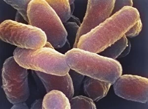
Background imageBacteria Collection: E. coli 0157: H7 bacteria

E. coli 0157: H7 bacteria
E. coli 0157:H7 bacteria. Coloured scanning electron micrograph (SEM) of Escherichia coli 0157:H7 bacteria, cause of foodborne illness

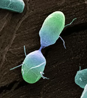
Background imageBacteria Collection: Salmonella bacterium dividing, SEM

Salmonella bacterium dividing, SEM
Salmonella bacterium dividing. Coloured scanning electron micrograph (SEM) of of a Salmonella bacterium dividing. The two new daughter cells are seen at upper right and lower left

Background imageBacteria Collection: Bacteria infecting a macrophage, SEM

Bacteria infecting a macrophage, SEM
Bacteria infecting a macrophage. Coloured scanning electron micrograph (SEM) of Mycobacterium tuberculosis bacteria (purple) infecting a macrophage white blood cell

Background imageBacteria Collection: False-colour TEM of Salmonella typhi

False-colour TEM of Salmonella typhi

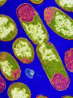
Background imageBacteria Collection: False-colour TEM of bacterium E. Coli

False-colour TEM of bacterium E. Coli
False colour transmission electron micrograph of the bacterium Escherichia coli, a normal member of human intestinal flora

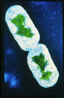
Background imageBacteria Collection: E. coli bacterium dividing

E. coli bacterium dividing

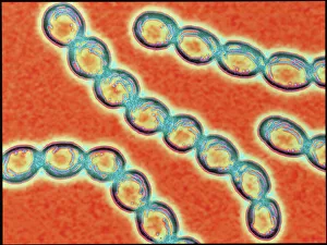
Background imageBacteria Collection: Chains of Streptococcus pyogenes bacteria

Chains of Streptococcus pyogenes bacteria

Background imageBacteria Collection: Psoriasis

Psoriasis
5855398 Psoriasis; (add.info.: Varieties of the autoimmune skin disease Psoriasis. Handcoloured copperplate stipple engraving by John Pass from John Wilkes' "Encyclopedia Londinensis,")

Background imageBacteria Collection: Measles

Measles
5855402 Measles; (add.info.: Measles, Rubeola vulgaris and R. nigra. Handcoloured copperplate stipple engraving by John Pass from John Wilkes' "Encyclopedia Londinensis," J)

Background imageBacteria Collection: Purple spots and rose rash

Purple spots and rose rash
5855400 Purple spots and rose rash; (add.info.: Skin diseases: Purple spots, Purpura simplex and P. haemorrhagica and rose rash, Exanthesis roseola)

Background imageBacteria Collection: Psoriasis

Psoriasis
5855396 Psoriasis; (add.info.: Skin diseases: Prurigo nodularis and Psoriasis varieties. (Lepriasis is an obsolete term for psoriasis and leprosy)

Background imageBacteria Collection: Skin diseases of infants

Skin diseases of infants
5855399 Skin diseases of infants; (add.info.: Red gum or Strophulus, a skin disease affecting infants, and Lichen simplex, a thickening of the skin after pruritus)

Background imageBacteria Collection: Scalp and skin diseases

Scalp and skin diseases
5855397 Scalp and skin diseases; (add.info.: Scalp diseases Porrigo crustacea and P. furfuracea, and skin diseases Ecthyma vulgaris and E)

Background imageBacteria Collection: Scarlet fever

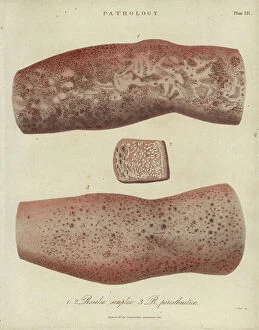
Scarlet fever
5855393 Scarlet fever; (add.info.: Scarlet fever, Rosalia simplex and R. paristhmitica. Handcoloured copperplate stipple engraving by John Pass from John Wilkes' "Encyclopedia)

Background imageBacteria Collection: Sycosis

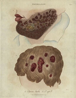
Sycosis
5855392 Sycosis; (add.info.: Sycosis barbae and Sycosis capilli, chronic inflammation of the hair follicles, especially of the beard and head)

Background imageBacteria Collection: Impetigo

Impetigo
5855391 Impetigo; (add.info.: Contagious skin diseases impetigo and rhypia. Handcoloured copperplate stipple engraving by John Pass from John Wilkes' "Encyclopedia Londinensis," J)

Background imageBacteria Collection: Scabies

Scabies
5855394 Scabies; (add.info.: Ulcerous skin disease Ecthyma cachecticum, and the seven-year itch, Scabies purulenta and S. exotica, caused by the Sarcoptes scabiei mite)

Background imageBacteria Collection: coronavirus

coronavirus
engin akyurt

Background imageBacteria Collection: coronavirus

coronavirus
engin akyurt

Background imageBacteria Collection: Petri Dish

Petri Dish
Thoth Adan

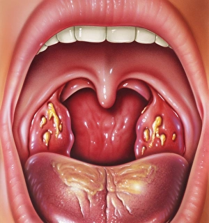
Background imageBacteria Collection: Tonsillitis

Tonsillitis. Artwork of the open mouth and throat of a patient with inflamed tonsils. They are flecked with infected yellow patches, due to tonsillitis

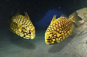
Background imageBacteria Collection: Picture No. 12479508

Picture No. 12479508
Pineapplefish, Cleidopus gloriamaris, inside Date:

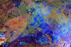
Background imageBacteria Collection: Bacteria (Leptothryx discophora) causing iridescent sheen on water surface with White willow leaf

Bacteria (Leptothryx discophora) causing iridescent sheen on water surface with White willow leaf (Salix alba). Sierra de Grazalema Natural Park, southern Spain, November

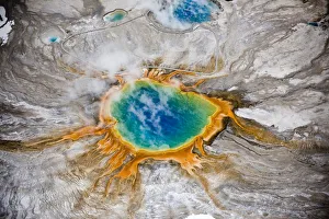
Background imageBacteria Collection: Aerial view of Grand Prismatic thermal spring in Yellowstone National Park, USA

Aerial view of Grand Prismatic thermal spring in Yellowstone National Park, USA. The bright colours are natural, coming from thermophile bacteria in the water

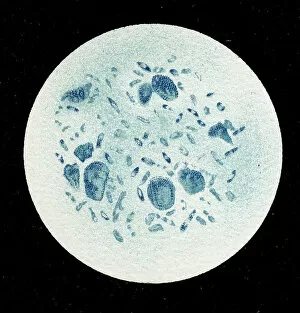
Background imageBacteria Collection: Colony of Haemophilus influenzae, 1906 (litho)

Colony of Haemophilus influenzae, 1906 (litho)
3501914 Colony of Haemophilus influenzae, 1906 (litho) by German School, (20th century); Private Collection; (add.info.: Bacteria constitute a large domain of prokaryotic microorganisms)

Background imageBacteria Collection: Colony of Mycobacterium leprae, 1906 (litho)

Colony of Mycobacterium leprae, 1906 (litho)
3501912 Colony of Mycobacterium leprae, 1906 (litho) by German School, (20th century); Private Collection; (add.info.: Bacteria constitute a large domain of prokaryotic microorganisms)

Background imageBacteria Collection: Colony of Micrococcus Gonorrhoea, 1906 (litho)

Colony of Micrococcus Gonorrhoea, 1906 (litho)
3501903 Colony of Micrococcus Gonorrhoea, 1906 (litho) by German School, (20th century); Private Collection; (add.info.: Bacteria constitute a large domain of prokaryotic microorganisms)

Background imageBacteria Collection: Colony of Streptococcus pneumoniae with and without bubble capsule, 1906 (litho)

Colony of Streptococcus pneumoniae with and without bubble capsule, 1906 (litho)
3501899 Colony of Streptococcus pneumoniae with and without bubble capsule, 1906 (litho) by German School, (20th century); Private Collection; (add.info)

Background imageBacteria Collection: Colony of Streptococcus Pneumoniae, 1906 (litho)

Colony of Streptococcus Pneumoniae, 1906 (litho)
3501898 Colony of Streptococcus Pneumoniae, 1906 (litho) by German School, (20th century); Private Collection; (add.info.: Bacteria constitute a large domain of prokaryotic microorganisms)

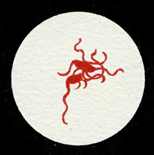
Background imageBacteria Collection: Spirillum of the Lyme disease causing bacteria, Spirochaetes Borrelia Recurrentis

Spirillum of the Lyme disease causing bacteria, Spirochaetes Borrelia Recurrentis, in blood sample, 1906 (litho)
3501919 Spirillum of the Lyme disease causing bacteria, Spirochaetes Borrelia Recurrentis, in blood sample, 1906 (litho) by German School

Background imageBacteria Collection: Colony of Vibrio cholerae, 1906 (litho)

Colony of Vibrio cholerae, 1906 (litho)
3501918 Colony of Vibrio cholerae, 1906 (litho) by German School, (20th century); Private Collection; (add.info.: Bacteria constitute a large domain of prokaryotic microorganisms)

Background imageBacteria Collection: Colony of Clostridium tetani with spores, 1906 (litho)

Colony of Clostridium tetani with spores, 1906 (litho)
3501913 Colony of Clostridium tetani with spores, 1906 (litho) by German School, (20th century); Private Collection; (add.info.: Bacteria constitute a large domain of prokaryotic microorganisms)

Background imageBacteria Collection: Colony of Mycobacterium tuberculosis, 1906 (litho)

Colony of Mycobacterium tuberculosis, 1906 (litho)
3501910 Colony of Mycobacterium tuberculosis, 1906 (litho) by German School, (20th century); Private Collection; (add.info.: Bacteria constitute a large domain of prokaryotic microorganisms)

Background imageBacteria Collection: A colony of Staphylococcus pyogenes after cell division, 1906 (litho)

A colony of Staphylococcus pyogenes after cell division, 1906 (litho)
3501897 A colony of Staphylococcus pyogenes after cell division, 1906 (litho) by German School, (20th century); Private Collection; (add.info)

Background imageBacteria Collection: Colony of Streptococcus pyogenes, 1906 (litho)

Colony of Streptococcus pyogenes, 1906 (litho)
3501894 Colony of Streptococcus pyogenes, 1906 (litho) by German School, (20th century); Private Collection; (add.info.: Bacteria constitute a large domain of prokaryotic microorganisms)

Background imageBacteria Collection: Varieties of microscopic Marine organisms, 1900 (litho)

Varieties of microscopic Marine organisms, 1900 (litho)
3501931 Varieties of microscopic Marine organisms, 1900 (litho) by German School, (20th century); Private Collection; Prismatic Pictures

Background imageBacteria Collection: Colony of Yersinia pestis, 1906 (litho)

Colony of Yersinia pestis, 1906 (litho)
3501917 Colony of Yersinia pestis, 1906 (litho) by German School, (20th century); Private Collection; (add.info.: Bacteria constitute a large domain of prokaryotic microorganisms)

Background imageBacteria Collection: Colony of Salmonella Typhi showing Bacilli with flagella, 1906 (litho)

Colony of Salmonella Typhi showing Bacilli with flagella, 1906 (litho)
3501916 Colony of Salmonella Typhi showing Bacilli with flagella, 1906 (litho) by German School, (20th century); Private Collection; (add.info)

Background imageBacteria Collection: Colony of Salmonella typhi, 1906 (litho)

Colony of Salmonella typhi, 1906 (litho)
3501915 Colony of Salmonella typhi, 1906 (litho) by German School, (20th century); Private Collection; (add.info.: Bacteria constitute a large domain of prokaryotic microorganisms)

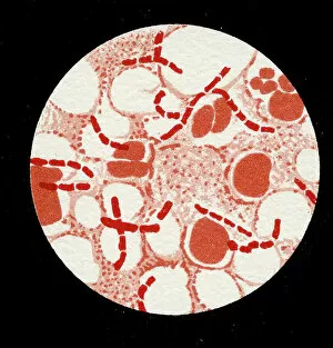
Background imageBacteria Collection: Colony of Bacillus Anthracis in a human spleen, 1906 (litho)

Colony of Bacillus Anthracis in a human spleen, 1906 (litho)
3501905 Colony of Bacillus Anthracis in a human spleen, 1906 (litho) by German School, (20th century); Private Collection; (add.info)

Background imageBacteria Collection: Spores of Bacillus Anthracis, 1906 (litho)

Spores of Bacillus Anthracis, 1906 (litho)
3501904 Spores of Bacillus Anthracis, 1906 (litho) by German School, (20th century); Private Collection; (add.info.: Bacteria constitute a large domain of prokaryotic microorganisms)

Background imageBacteria Collection: Colony of Micrococcus Gonorrhoea found in the Urethra, 1906 (litho)

Colony of Micrococcus Gonorrhoea found in the Urethra, 1906 (litho)
3501900 Colony of Micrococcus Gonorrhoea found in the Urethra, 1906 (litho) by German School, (20th century); Private Collection; (add.info)

Background imageBacteria Collection: Colony of Corynebacterium Diphtheriae, 1906 (litho)

Colony of Corynebacterium Diphtheriae, 1906 (litho)
3501907 Colony of Corynebacterium Diphtheriae, 1906 (litho) by German School, (20th century); Private Collection; (add.info.: Bacteria constitute a large domain of prokaryotic microorganisms)

Background imageBacteria Collection: Colony of Bacterium mallei, 1906 (litho)

Colony of Bacterium mallei, 1906 (litho)
3501906 Colony of Bacterium mallei, 1906 (litho) by German School, (20th century); Private Collection; (add.info.: Bacteria constitute a large domain of prokaryotic microorganisms)

Background imageBacteria Collection: Staphylococcus colony, 1905 (litho)

Staphylococcus colony, 1905 (litho)
3501895 Staphylococcus colony, 1905 (litho) by German School, (20th century); Private Collection; (add.info.: Bacteria constitute a large domain of prokaryotic microorganisms)

Background imageBacteria Collection: Colony of Mycobacterium Tuberculosis, 1906 (litho)

Colony of Mycobacterium Tuberculosis, 1906 (litho)
3501911 Colony of Mycobacterium Tuberculosis, 1906 (litho) by German School, (20th century); Private Collection; (add.info.: Bacteria constitute a large domain of prokaryotic microorganisms)

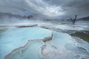
Background imageBacteria Collection: Deposits of travertine colored by thermophilic bacteria, Upper Terraces Mammoth Hot Springs

Deposits of travertine colored by thermophilic bacteria, Upper Terraces Mammoth Hot Springs, Yellowstone National Park. Date: 21-09-2019

Background imageBacteria Collection: Soil bacteria cultured on nutrient agar from a soil sample collected in Via Gellia Woods, Derbyshire

Soil bacteria cultured on nutrient agar from a soil sample collected in Via Gellia Woods, Derbyshire. Image size in real life 10mm across. UK. March


All Professionally Made to Order for Quick Shipping

"Bacteria: The Microscopic Marvels of Yellowstone National Park" In the heart of Wyoming, USA lies the breathtaking Yellowstone National Park, a haven for nature enthusiasts and scientists alike. Amidst its natural wonders, one can find an extraordinary display of microbial life at the Grand Prismatic Spring in the Midway Geyser Basin. Picture No. 11675590 captures this mesmerizing sight, showcasing vibrant hues that seem to dance upon the water's surface. However, what may appear as an artistic masterpiece is actually a thriving community of bacteria. Zooming in closer with SEM C018 / 8596 reveals a neutrophil engulfing MRSA - Methicillin-resistant Staphylococcus aureus. This image serves as a reminder of how these tiny organisms can pose significant threats to human health. Another SEM image showcases E. Coli bacteria, reminding us that not all it can harmful; some play crucial roles in our digestive system and even aid scientific research. Moving back to Yellowstone's enchantment, we encounter Salmonella bacteria through another captivating SEM photograph. These microscopic creatures serve as a stark reminder that even within such serene landscapes, dangers lurk unseen. Coloured TEM imagery introduces us to Yersinia pestis bacteria – responsible for causing devastating outbreaks like the infamous Black Death throughout history. Such visuals emphasize their intricate structures and highlight why understanding them is vital for public health efforts. Venturing beyond real-life images into computer artwork brings forth a beta DNA segment surrounded by spheres—a representation of genetic material essential for bacterial survival and reproduction. Artistic depictions also shed light on various cell types found within these microorganisms' complex world—each playing unique roles necessary for their survival and proliferation. Historical diagrams reveal cultures of Anthrax—an infectious disease with severe consequences if left unchecked—underscoring humanity's ongoing battle against bacterial infections throughout time. Tuberculosis bacteria make their appearance too—a haunting reminder that despite medical advancements, infectious diseases continue to challenge us.