
Arteries Collection (page 6)
Arteries, the lifelines of our bodies, play a crucial role in maintaining our overall health and well-being


All Professionally Made to Order for Quick Shipping
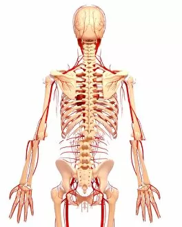
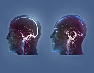
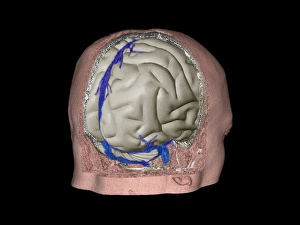
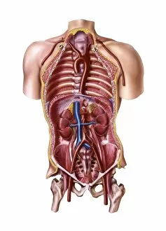
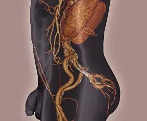
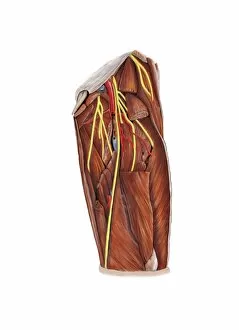
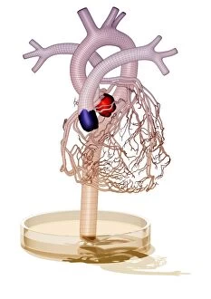
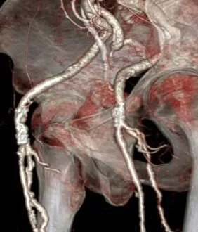
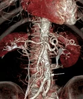
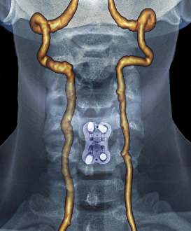
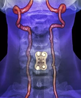
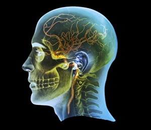
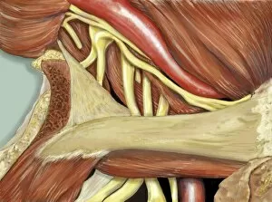
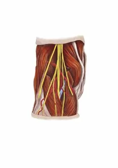
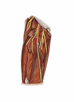
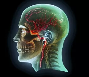
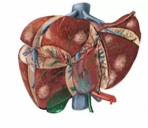
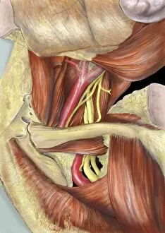
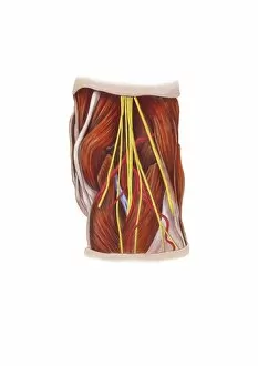
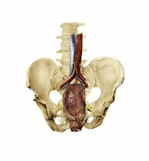
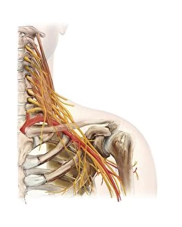
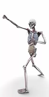
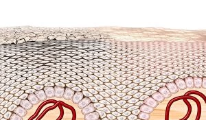
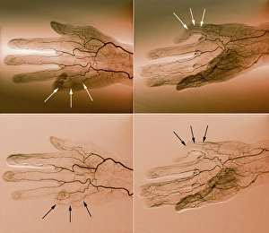
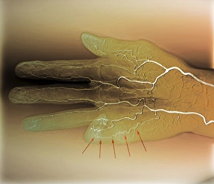
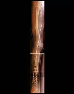
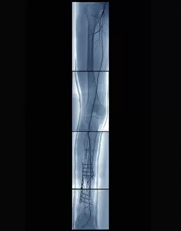
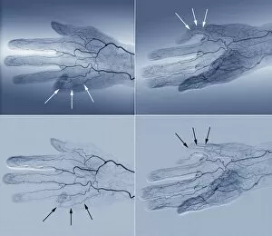
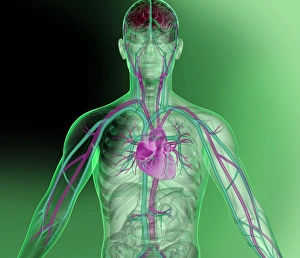
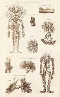
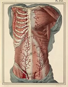
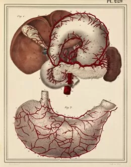
Arteries, the lifelines of our bodies, play a crucial role in maintaining our overall health and well-being. These intricate blood vessels are responsible for carrying oxygen-rich blood from the heart to every part of our body, ensuring that all organs receive the nutrients they need to function properly. In the realm of medical imaging, advancements like 3D angiograms have revolutionized our understanding of arteries. With technologies such as C007/1981 scans, we can now visualize these brain blood vessels in unprecedented detail. Similarly, full-body scans and MRI scans provide comprehensive insights into the complex network intertwined with bones throughout the human body. The cardiovascular system is an awe-inspiring masterpiece that has captivated artists throughout history. Historical artwork showcases their fascination with depicting the heart alongside veins and arteries - a testament to its vital importance in sustaining life. From 18th-century depictions of arterial systems to 19th-century illustrations exploring neck vascular anatomy, these artworks beautifully capture both scientific knowledge and artistic expression. As we delve deeper into understanding artery anatomy, it becomes clear how intricately woven this network truly is. Neck anatomy artwork from centuries ago offers glimpses into early attempts at comprehending this complex system within our bodies. Human heart anatomical drawings further highlight how artists have sought to portray this essential organ accurately. Ultimately, when contemplating arteries' significance within medical science or appreciating their beauty through artistry, one cannot overlook their pivotal role in maintaining good health. The study of these blood vessels remains integral to medical research and practice today – reminding us just how remarkable our bodies truly are.