
Aortic Valve Collection
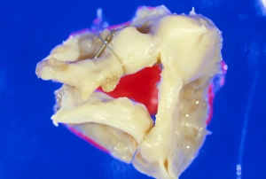
The aortic valve, an integral part of the human heart's anatomy, plays a crucial role in maintaining proper blood flow


All Professionally Made to Order for Quick Shipping
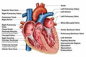
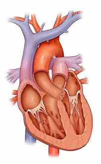
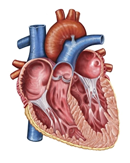
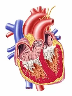
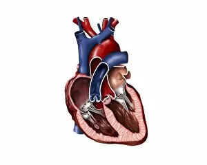
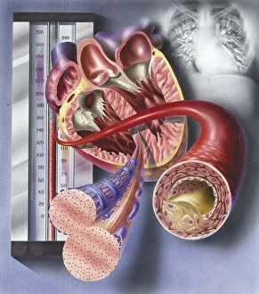
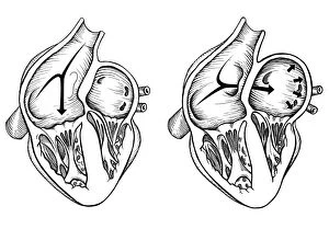
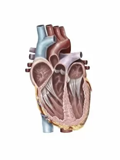
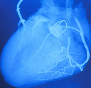
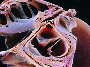
The aortic valve, an integral part of the human heart's anatomy, plays a crucial role in maintaining proper blood flow. Located within the interior of the heart, specifically in the frontal section and cross section, this valve ensures efficient circulation throughout the body. With its intricate design and function, it regulates blood flow from the left ventricle into the aorta. When examining a normal heart's cross section or internal view, one can observe how the aortic valve works alongside other valves such as pulmonary valve, mitral valve, and tricuspid. These valves collectively ensure that blood flows smoothly through different chambers of the heart. However, complications may arise when abnormalities occur within these valves or structures surrounding them. For instance, comparing a normal heart to one with a patent foramen ovale reveals distinct differences in their functionality. Such comparisons aid medical professionals in diagnosing conditions related to these vital components. Furthermore, an enlarged left ventricle can also impact how well the aortic valve functions. This condition often occurs due to various factors like high blood pressure or cardiovascular diseases. Understanding and studying every aspect of our hearts' interior is essential for maintaining overall health and preventing potential issues related to cardiac function. The intricate details of muscle cells and even pathologies like an atherosclerotic artery provide valuable insights into how our hearts work on both microscopic and macroscopic levels. Exploring images depicting different aspects of human hearts – be it anatomical sections or internal views – helps us appreciate just how remarkable our bodies are while emphasizing why we must prioritize cardiovascular health.